6K2Z

Human Galectin-14 with lactose


Experimental Data Snapshot

  • Method: X-RAY DIFFRACTION
  • Resolution: 2.00 Å
  • R-Value Free: 0.246 
  • R-Value Work: 0.202 
  • R-Value Observed: 0.206 

wwPDB Validation   3D Report Full Report


This is version 2.2 of the entry. See complete history


Literature

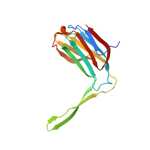
Structure-function studies of galectin-14, an important effector molecule in embryology.

Si, Y.Li, Y.Yang, T.Li, X.Ayala, G.J.Mayo, K.H.Tai, G.Su, J.Zhou, Y.

(2021) FEBS J 288: 1041-1055

  • DOI: https://doi.org/10.1111/febs.15441
  • Primary Citation of Related Structures:  
    6K2Y, 6K2Z

  • PubMed Abstract: 

    The expression of prototype galectin-14 (Gal-14) in human placenta is higher than any other galectin, suggesting that it may play a role in fetal development and regulation of immune tolerance during pregnancy. Here, we solved the crystal structure of dimeric Gal-14 and found that its global fold is significantly different from that of other galectins with two β-strands (S5 and S6) extending from one monomer and contributing to the carbohydrate-binding domain of the other. The hemagglutination assay showed that this lectin could induce agglutination of chicken erythrocytes, even though lactose could not inhibit Gal-14-induced agglutination activity. Calorimetry indicates that lactose does not interact with this lectin. Compared to galectin-1, galectin-3, and galectin-8, Gal-14 has two key amino acids (a histidine and an arginine) in the normally conserved, canonical sugar-binding site, which are substituted by glutamine (Gln53) and histidine (His57), thus likely explaining why lactose binding to this lectin is very weak. Lactose was observed in the ligand-binding site of one Gal-14 structure, most likely because ligand binding is weak and crystals were allowed to grow over a long period of time in the presence of lactose. We also found that EGFP-tagged Gal-14 is primarily localized within the nucleus of different cell types. In addition, Gal-14 colocalized with c-Rel (a member of NF-κB family) in HeLa cells. These findings indicate that Gal-14 might regulate signal transduction pathways through NF-κB hubs. Overall, the present study provides impetus for further research into the function of Gal-14 in embryology.


  • Organizational Affiliation

    Engineering Research Center of Glycoconjugates Ministry of Education, Jilin Provincial Key Laboratory of Chemistry and Biology of Changbai Mountain Natural Drugs, School of Life Sciences, Northeast Normal University, Changchun, China.


Macromolecules
Find similar proteins by:  (by identity cutoff)  |  3D Structure
Entity ID: 1
MoleculeChains Sequence LengthOrganismDetailsImage
Placental protein 13-like
A, B
142Homo sapiensMutation(s): 0 
Gene Names: LGALS14PPL13
UniProt & NIH Common Fund Data Resources
Find proteins for Q8TCE9 (Homo sapiens)
Explore Q8TCE9 
Go to UniProtKB:  Q8TCE9
PHAROS:  Q8TCE9
GTEx:  ENSG00000006659 
Entity Groups  
Sequence Clusters30% Identity50% Identity70% Identity90% Identity95% Identity100% Identity
UniProt GroupQ8TCE9
Sequence Annotations
Expand
  • Reference Sequence
Oligosaccharides

Help

Entity ID: 2
MoleculeChains Length2D Diagram Glycosylation3D Interactions
beta-D-galactopyranose-(1-4)-beta-D-glucopyranose
C
2N/A
Glycosylation Resources
GlyTouCan:  G84224TW
GlyCosmos:  G84224TW
GlyGen:  G84224TW
Entity ID: 3
MoleculeChains Length2D Diagram Glycosylation3D Interactions
beta-D-galactopyranose-(1-4)-alpha-D-glucopyranose
D
2N/A
Glycosylation Resources
GlyTouCan:  G88362QR
GlyCosmos:  G88362QR
GlyGen:  G88362QR
Biologically Interesting Molecules (External Reference) 2 Unique
Experimental Data & Validation

Experimental Data

  • Method: X-RAY DIFFRACTION
  • Resolution: 2.00 Å
  • R-Value Free: 0.246 
  • R-Value Work: 0.202 
  • R-Value Observed: 0.206 
  • Space Group: P 21 21 21
Unit Cell:
Length ( Å )Angle ( ˚ )
a = 35.813α = 90
b = 63.354β = 90
c = 130.879γ = 90
Software Package:
Software NamePurpose
PHENIXrefinement

Structure Validation

View Full Validation Report


Entry History 

Deposition Data

  • Released Date: 2020-06-17 
  • Deposition Author(s): Su, J.

Revision History  (Full details and data files)

  • Version 1.0: 2020-06-17
    Type: Initial release
  • Version 1.1: 2020-06-24
    Changes: Database references
  • Version 2.0: 2020-07-29
    Type: Remediation
    Reason: Carbohydrate remediation
    Changes: Atomic model, Data collection, Derived calculations, Structure summary
  • Version 2.1: 2021-03-10
    Changes: Database references, Derived calculations, Structure summary
  • Version 2.2: 2024-03-27
    Changes: Data collection, Database references