6K24

Structure of the Rhodium Mesoporphyrin IX-Reconstituted CYP102A1 Haem Domain with N-Abietoyl-L-Tryptophan


Experimental Data Snapshot

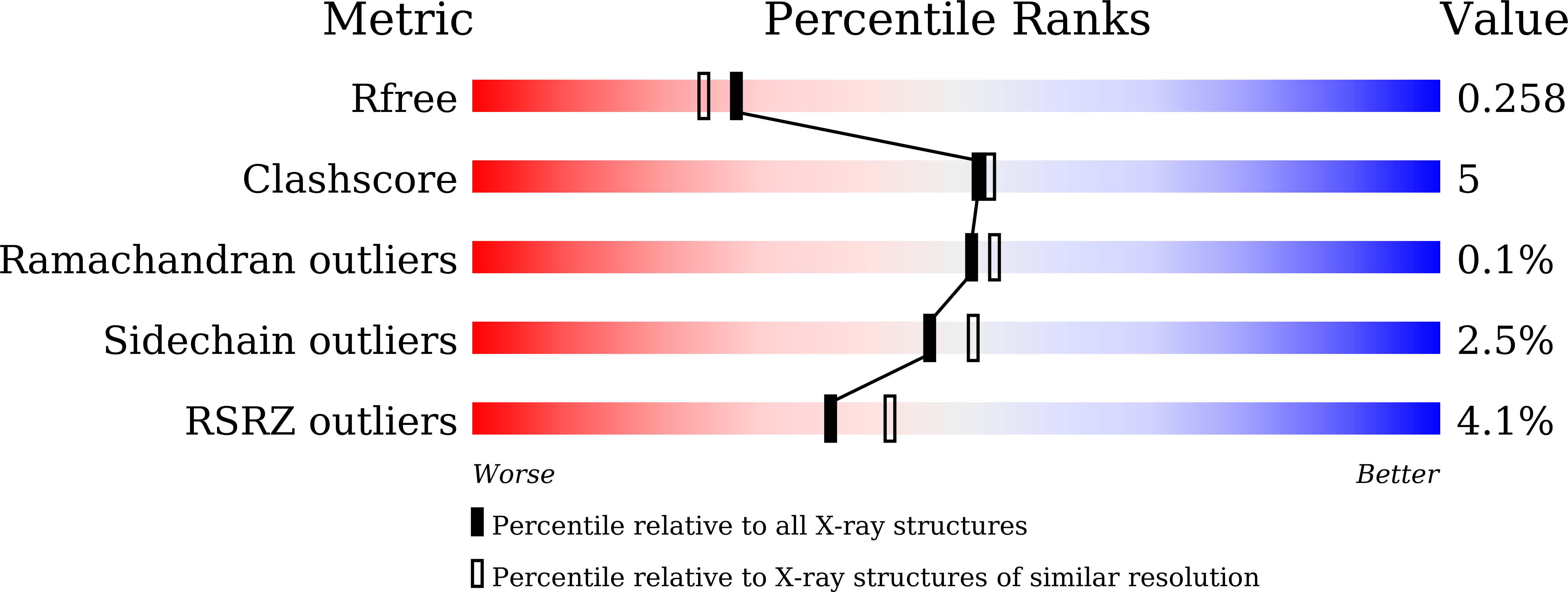
  • Method: X-RAY DIFFRACTION
  • Resolution: 2.10 Å
  • R-Value Free: 0.256 
  • R-Value Work: 0.208 
  • R-Value Observed: 0.211 

wwPDB Validation   3D Report Full Report


Ligand Structure Quality Assessment 


This is version 1.3 of the entry. See complete history


Literature

Crystals in Minutes: Instant On-Site Microcrystallisation of Various Flavours of the CYP102A1 (P450BM3) Haem Domain.

Stanfield, J.K.Omura, K.Matsumoto, A.Kasai, C.Sugimoto, H.Shiro, Y.Watanabe, Y.Shoji, O.

(2020) Angew Chem Int Ed Engl 59: 7611-7618

  • DOI: https://doi.org/10.1002/anie.201913407
  • Primary Citation of Related Structures:  
    6JLV, 6JMH, 6JMW, 6JO1, 6JS8, 6JVC, 6JZS, 6K24, 6K58, 6K9S

  • PubMed Abstract: 

    Despite CYP102A1 (P450BM3) representing one of the most extensively researched metalloenzymes, crystallisation of its haem domain upon modification can be a challenge. Crystal structures are indispensable for the efficient structure-based design of P450BM3 as a biocatalyst. The abietane diterpenoid derivative N-abietoyl-l-tryptophan (AbiATrp) is an outstanding crystallisation accelerator for the wild-type P450BM3 haem domain, with visible crystals forming within 2 hours and diffracting to a near-atomic resolution of 1.22 Å. Using these crystals as seeds in a cross-microseeding approach, an assortment of P450BM3 haem domain crystal structures, containing previously uncrystallisable decoy molecules and diverse artificial metalloporphyrins binding various ligand molecules, as well as heavily tagged haem-domain variants, could be determined. Some of the structures reported herein could be used as models of different stages of the P450BM3 catalytic cycle.


  • Organizational Affiliation

    Department of Chemistry, Graduate School of Science, Nagoya University, Furo-cho, Chikusa-ku, Nagoya, 464-8602, Japan.


Macromolecules
Find similar proteins by:  (by identity cutoff)  |  3D Structure
Entity ID: 1
MoleculeChains Sequence LengthOrganismDetailsImage
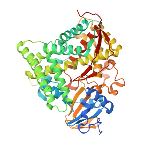
Bifunctional cytochrome P450/NADPH--P450 reductase
A, B
481Priestia megateriumMutation(s): 0 
Gene Names: cyp102A1
EC: 1.14.14.1 (PDB Primary Data), 1.6.2.4 (PDB Primary Data)
UniProt
Find proteins for P14779 (Priestia megaterium (strain ATCC 14581 / DSM 32 / CCUG 1817 / JCM 2506 / NBRC 15308 / NCIMB 9376 / NCTC 10342 / NRRL B-14308 / VKM B-512 / Ford 19))
Explore P14779 
Go to UniProtKB:  P14779
Entity Groups  
Sequence Clusters30% Identity50% Identity70% Identity90% Identity95% Identity100% Identity
UniProt GroupP14779
Sequence Annotations
Expand
  • Reference Sequence
Experimental Data & Validation

Experimental Data

  • Method: X-RAY DIFFRACTION
  • Resolution: 2.10 Å
  • R-Value Free: 0.256 
  • R-Value Work: 0.208 
  • R-Value Observed: 0.211 
  • Space Group: P 1 21 1
Unit Cell:
Length ( Å )Angle ( ˚ )
a = 59.082α = 90
b = 149.182β = 99.76
c = 65.213γ = 90
Software Package:
Software NamePurpose
REFMACrefinement
XDSdata reduction
Aimlessdata scaling
MOLREPphasing
PDB_EXTRACTdata extraction

Structure Validation

View Full Validation Report


Ligand Structure Quality Assessment 


Entry History & Funding Information

Deposition Data


Funding OrganizationLocationGrant Number
Japan Science and TechnologyJapanJPMJCR15P3

Revision History  (Full details and data files)

  • Version 1.0: 2020-03-18
    Type: Initial release
  • Version 1.1: 2020-03-25
    Changes: Database references
  • Version 1.2: 2020-05-06
    Changes: Database references
  • Version 1.3: 2023-11-22
    Changes: Data collection, Database references, Derived calculations, Refinement description, Structure summary