6JM4

The crystal structure of PB1 homo-dimer of human P62/SQSTM1


Experimental Data Snapshot

  • Method: X-RAY DIFFRACTION
  • Resolution: 3.20 Å
  • R-Value Free: 0.276 
  • R-Value Work: 0.239 
  • R-Value Observed: 0.243 

wwPDB Validation   3D Report Full Report


This is version 1.1 of the entry. See complete history


Literature

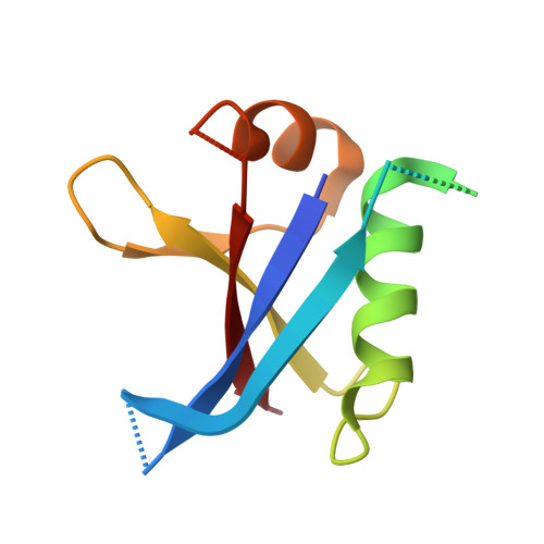
Oligomer Model of PB1 Domain of p62/SQSTM1 Based on Crystal Structure of Homo-Dimer and Calculation of Helical Characteristics.

Lim, D.Lee, H.S.Ku, B.Shin, H.C.Kim, S.J.

(2019) Mol Cells 42: 729-738

  • DOI: https://doi.org/10.14348/molcells.2019.0096
  • Primary Citation of Related Structures:  
    6JM4

  • PubMed Abstract: 

    Autophagy is an important process for protein recycling. Oligomerization of p62/SQSTM1 is an essential step in this process and is achieved in two steps. Phox and Bem1p (PB1) domains can oligomerize through both basic and acidic surfaces in each molecule. The ZZ-type zinc finger (ZZ) domain binds to target proteins and promotes higheroligomerization of p62. This mechanism is an important step in routing target proteins to the autophagosome. Here, we determined the crystal structure of the PB1 homo-dimer and modeled the p62 PB1 oligomers. These oligomer models were represented by a cylindrical helix and were compared with the previously determined electron microscopic map of a PB1 oligomer. To accurately compare, we mathematically calculated the lead length and radius of the helical oligomers. Our PB1 oligomer model fits the electron microscopy map and is both bendable and stretchable as a flexible helical filament.


  • Organizational Affiliation

    Disease Target Structure Research Center, Korea Research Institute of Bioscience and Biotechnology, Daejeon 34141, Korea.


Macromolecules
Find similar proteins by:  (by identity cutoff)  |  3D Structure
Entity ID: 1
MoleculeChains Sequence LengthOrganismDetailsImage
Sequestosome-1A,
D [auth C]
102Homo sapiensMutation(s): 4 
Gene Names: SQSTM1ORCAOSIL
UniProt & NIH Common Fund Data Resources
Find proteins for Q13501 (Homo sapiens)
Explore Q13501 
Go to UniProtKB:  Q13501
PHAROS:  Q13501
GTEx:  ENSG00000161011 
Entity Groups  
Sequence Clusters30% Identity50% Identity70% Identity90% Identity95% Identity100% Identity
UniProt GroupQ13501
Sequence Annotations
Expand
  • Reference Sequence
Find similar proteins by:  (by identity cutoff)  |  3D Structure
Entity ID: 2
MoleculeChains Sequence LengthOrganismDetailsImage
Sequestosome-1B,
C [auth D]
102Homo sapiensMutation(s): 3 
Gene Names: SQSTM1ORCAOSIL
UniProt & NIH Common Fund Data Resources
Find proteins for Q13501 (Homo sapiens)
Explore Q13501 
Go to UniProtKB:  Q13501
PHAROS:  Q13501
GTEx:  ENSG00000161011 
Entity Groups  
Sequence Clusters30% Identity50% Identity70% Identity90% Identity95% Identity100% Identity
UniProt GroupQ13501
Sequence Annotations
Expand
  • Reference Sequence
Experimental Data & Validation

Experimental Data

  • Method: X-RAY DIFFRACTION
  • Resolution: 3.20 Å
  • R-Value Free: 0.276 
  • R-Value Work: 0.239 
  • R-Value Observed: 0.243 
  • Space Group: C 1 2 1
Unit Cell:
Length ( Å )Angle ( ˚ )
a = 112.483α = 90
b = 53.517β = 94.287
c = 61.457γ = 90
Software Package:
Software NamePurpose
PHENIXrefinement
HKL-2000data reduction
HKL-2000data scaling
PHASERphasing

Structure Validation

View Full Validation Report


Entry History & Funding Information

Deposition Data


Funding OrganizationLocationGrant Number
National Research Foundation (Korea)Korea, Republic Of2015M3A9B5030308
Ministry of Science, ICT and Future PlanningKorea, Republic Of--

Revision History  (Full details and data files)

  • Version 1.0: 2020-01-15
    Type: Initial release
  • Version 1.1: 2023-11-22
    Changes: Data collection, Database references, Refinement description