6IXN

Crystal structure of isocitrate dehydrogenase from Ostreococcus tauri in complex with NAD+ and citrate


Experimental Data Snapshot

  • Method: X-RAY DIFFRACTION
  • Resolution: 1.87 Å
  • R-Value Free: 0.222 
  • R-Value Work: 0.195 
  • R-Value Observed: 0.197 

wwPDB Validation   3D Report Full Report


Ligand Structure Quality Assessment 


This is version 1.2 of the entry. See complete history


Literature

Crystal structures of NAD + -linked isocitrate dehydrogenase from the green alga Ostreococcus tauri and its evolutionary relationship with eukaryotic NADP + -linked homologs.

Tang, W.Wu, M.Qin, N.Liu, L.Meng, R.Wang, C.Wang, P.Zang, J.Zhu, G.

(2021) Arch Biochem Biophys 708: 108898-108898

  • DOI: https://doi.org/10.1016/j.abb.2021.108898
  • Primary Citation of Related Structures:  
    6IXL, 6IXN, 6IXT, 7E2W

  • PubMed Abstract: 

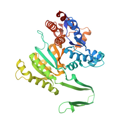
    NAD + -linked isocitrate dehydrogenases (NAD-IDHs) catalyze the oxidative decarboxylation of isocitrate into α-ketoglutarate. Previously, we identified a novel phylogenetic clade including NAD-IDHs from several algae in the type II subfamily, represented by homodimeric NAD-IDH from Ostreococcus tauri (OtIDH). However, due to its lack of a crystalline structure, the molecular mechanisms of the ligand binding and catalysis of OtIDH are little known. Here, we elucidate four high-resolution crystal structures of OtIDH in a ligand-free and various ligand-bound forms that capture at least three states in the catalytic cycle: open, semi-closed, and fully closed. Our results indicate that OtIDH shows several novel interactions with NAD + , unlike type I NAD-IDHs, as well as a strictly conserved substrate binding mode that is similar to other homologs. The central roles of Lys283' in dual coenzyme recognition and Lys234 in catalysis were also revealed. In addition, the crystal structures obtained here also allow us to understand the catalytic mechanism. As expected, structural comparisons reveal that OtIDH has a very high structural similarity to eukaryotic NADP + -linked IDHs (NADP-IDHs) within the type II subfamily rather than with the previously reported NAD-IDHs within the type I subfamily. It has also been demonstrated that OtIDH exhibits substantial conformation changes upon ligand binding, similar to eukaryotic NADP-IDHs. These results unambiguously support our hypothesis that OtIDH and OtIDH-like homologs are possible evolutionary ancestors of eukaryotic NADP-IDHs in type II subfamily.


  • Organizational Affiliation

    Anhui Provincial Key Laboratory of Molecular Enzymology and Mechanism of Major Diseases and Key Laboratory of Biomedicine in Gene Diseases and Health of Anhui Higher Education Institutes, No.1 Beijing East Road, College of Life Sciences, Anhui Normal University, Wuhu, Anhui, 241000, China; Department of Biochemistry and Molecular Biology, School of Laboratory Medicine, Bengbu Medical College, Bengbu, Anhui, 233030, China.


Macromolecules
Find similar proteins by:  (by identity cutoff)  |  3D Structure
Entity ID: 1
MoleculeChains Sequence LengthOrganismDetailsImage
Isocitrate dehydrogenase
A, B, C, D
418Ostreococcus tauriMutation(s): 0 
Gene Names: BE221DRAFT_192402
UniProt
Find proteins for A0A096P8D3 (Ostreococcus tauri)
Explore A0A096P8D3 
Go to UniProtKB:  A0A096P8D3
Entity Groups  
Sequence Clusters30% Identity50% Identity70% Identity90% Identity95% Identity100% Identity
UniProt GroupA0A096P8D3
Sequence Annotations
Expand
  • Reference Sequence
Small Molecules
Ligands 5 Unique
IDChains Name / Formula / InChI Key2D Diagram3D Interactions
NAD
Query on NAD

Download Ideal Coordinates CCD File 
I [auth A],
O [auth B],
T [auth C],
Z [auth D]
NICOTINAMIDE-ADENINE-DINUCLEOTIDE
C21 H27 N7 O14 P2
BAWFJGJZGIEFAR-NNYOXOHSSA-N
FLC
Query on FLC

Download Ideal Coordinates CCD File 
H [auth A],
N [auth B],
S [auth C],
Y [auth D]
CITRATE ANION
C6 H5 O7
KRKNYBCHXYNGOX-UHFFFAOYSA-K
PEG
Query on PEG

Download Ideal Coordinates CCD File 
M [auth B]DI(HYDROXYETHYL)ETHER
C4 H10 O3
MTHSVFCYNBDYFN-UHFFFAOYSA-N
SO4
Query on SO4

Download Ideal Coordinates CCD File 
E [auth A]
F [auth A]
J [auth B]
K [auth B]
P [auth C]
E [auth A],
F [auth A],
J [auth B],
K [auth B],
P [auth C],
Q [auth C],
U [auth D],
V [auth D]
SULFATE ION
O4 S
QAOWNCQODCNURD-UHFFFAOYSA-L
GOL
Query on GOL

Download Ideal Coordinates CCD File 
G [auth A],
L [auth B],
R [auth C],
W [auth D],
X [auth D]
GLYCEROL
C3 H8 O3
PEDCQBHIVMGVHV-UHFFFAOYSA-N
Experimental Data & Validation

Experimental Data

  • Method: X-RAY DIFFRACTION
  • Resolution: 1.87 Å
  • R-Value Free: 0.222 
  • R-Value Work: 0.195 
  • R-Value Observed: 0.197 
  • Space Group: P 1 21 1
Unit Cell:
Length ( Å )Angle ( ˚ )
a = 103.115α = 90
b = 102.928β = 104.07
c = 103.29γ = 90
Software Package:
Software NamePurpose
REFMACrefinement
XDSdata reduction
SCALAdata scaling
MOLREPphasing

Structure Validation

View Full Validation Report


Ligand Structure Quality Assessment 


Entry History & Funding Information

Deposition Data


Funding OrganizationLocationGrant Number
National Natural Science Foundation of ChinaChina31570010

Revision History  (Full details and data files)

  • Version 1.0: 2019-12-18
    Type: Initial release
  • Version 1.1: 2021-06-16
    Changes: Database references
  • Version 1.2: 2024-03-27
    Changes: Data collection, Database references