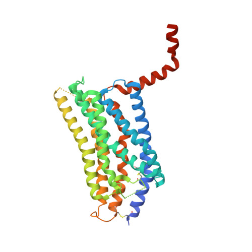
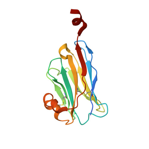
6IBB

Crystal structure of the rat isoform of the succinate receptor SUCNR1 (GPR91) in complex with a nanobody


Experimental Data Snapshot

  • Method: X-RAY DIFFRACTION
  • Resolution: 2.12 Å
  • R-Value Free: 0.216 
  • R-Value Work: 0.178 
  • R-Value Observed: 0.180 

wwPDB Validation   3D Report Full Report


Ligand Structure Quality Assessment 


This is version 1.3 of the entry. See complete history


Literature

Structural basis of species-selective antagonist binding to the succinate receptor.

Haffke, M.Fehlmann, D.Rummel, G.Boivineau, J.Duckely, M.Gommermann, N.Cotesta, S.Sirockin, F.Freuler, F.Littlewood-Evans, A.Kaupmann, K.Jaakola, V.P.

(2019) Nature 574: 581-585

  • DOI: https://doi.org/10.1038/s41586-019-1663-8
  • Primary Citation of Related Structures:  
    6IBB, 6RNK

  • PubMed Abstract: 

    The tricarboxylic acid cycle intermediate succinate is involved in metabolic processes and plays a crucial role in the homeostasis of mitochondrial reactive oxygen species 1 . The receptor responsible for succinate signalling, SUCNR1 (also known as GPR91), is a member of the G-protein-coupled-receptor family 2 and links succinate signalling to renin-induced hypertension, retinal angiogenesis and inflammation 3-5 . Because SUCNR1 senses succinate as an immunological danger signal 6 -which has relevance for diseases including ulcerative colitis, liver fibrosis 7 , diabetes and rheumatoid arthritis 3,8 -it is of interest as a therapeutic target. Here we report the high-resolution crystal structure of rat SUCNR1 in complex with an intracellular binding nanobody in the inactive conformation. Structure-based mutagenesis and radioligand-binding studies, in conjunction with molecular modelling, identified key residues for species-selective antagonist binding and enabled the determination of the high-resolution crystal structure of a humanized rat SUCNR1 in complex with a high-affinity, human-selective antagonist denoted NF-56-EJ40. We anticipate that these structural insights into the architecture of the succinate receptor and its antagonist selectivity will enable structure-based drug discovery and will further help to elucidate the function of SUCNR1 in vitro and in vivo.


  • Organizational Affiliation

    Chemical Biology & Therapeutics, Novartis Institutes for BioMedical Research, Novartis Pharma AG, Basel, Switzerland. matthias.haffke@novartis.com.


Macromolecules
Find similar proteins by:  (by identity cutoff)  |  3D Structure
Entity ID: 1
MoleculeChains Sequence LengthOrganismDetailsImage
Succinate receptor 1
A, C
342Rattus norvegicusMutation(s): 0 
Gene Names: Sucnr1Gpr91
Membrane Entity: Yes 
UniProt
Find proteins for Q6IYF9 (Rattus norvegicus)
Explore Q6IYF9 
Go to UniProtKB:  Q6IYF9
Entity Groups  
Sequence Clusters30% Identity50% Identity70% Identity90% Identity95% Identity100% Identity
UniProt GroupQ6IYF9
Sequence Annotations
Expand
  • Reference Sequence
Find similar proteins by:  (by identity cutoff)  |  3D Structure
Entity ID: 2
MoleculeChains Sequence LengthOrganismDetailsImage
Nanobody6
B, D
142Vicugna pacosMutation(s): 0 
Entity Groups  
Sequence Clusters30% Identity50% Identity70% Identity90% Identity95% Identity100% Identity
Sequence Annotations
Expand
  • Reference Sequence
Small Molecules
Ligands 4 Unique
IDChains Name / Formula / InChI Key2D Diagram3D Interactions
CLR
Query on CLR

Download Ideal Coordinates CCD File 
Q [auth C]CHOLESTEROL
C27 H46 O
HVYWMOMLDIMFJA-DPAQBDIFSA-N
OLC
Query on OLC

Download Ideal Coordinates CCD File 
E [auth A]
F [auth A]
G [auth A]
H [auth A]
I [auth A]
E [auth A],
F [auth A],
G [auth A],
H [auth A],
I [auth A],
J [auth A],
K [auth A],
L [auth A],
O [auth C],
P [auth C],
R [auth C],
S [auth C],
T [auth C],
U [auth C],
V [auth C]
(2R)-2,3-dihydroxypropyl (9Z)-octadec-9-enoate
C21 H40 O4
RZRNAYUHWVFMIP-GDCKJWNLSA-N
H95
Query on H95

Download Ideal Coordinates CCD File 
N [auth A](2~{S},5~{R})-hexane-2,5-diol
C6 H14 O2
OHMBHFSEKCCCBW-OLQVQODUSA-N
GOL
Query on GOL

Download Ideal Coordinates CCD File 
M [auth A],
W [auth C]
GLYCEROL
C3 H8 O3
PEDCQBHIVMGVHV-UHFFFAOYSA-N
Experimental Data & Validation

Experimental Data

  • Method: X-RAY DIFFRACTION
  • Resolution: 2.12 Å
  • R-Value Free: 0.216 
  • R-Value Work: 0.178 
  • R-Value Observed: 0.180 
  • Space Group: P 1 21 1
Unit Cell:
Length ( Å )Angle ( ˚ )
a = 60.22α = 90
b = 164β = 102.63
c = 63.42γ = 90
Software Package:
Software NamePurpose
BUSTERrefinement
XSCALEdata scaling
PHASERphasing
PDB_EXTRACTdata extraction
XDSdata reduction

Structure Validation

View Full Validation Report


Ligand Structure Quality Assessment 


Entry History 

Deposition Data

Revision History  (Full details and data files)

  • Version 1.0: 2019-08-14
    Type: Initial release
  • Version 1.1: 2019-10-16
    Changes: Data collection, Database references
  • Version 1.2: 2019-11-06
    Changes: Data collection, Database references
  • Version 1.3: 2024-01-24
    Changes: Data collection, Database references, Refinement description