6I1A

Crystal structure of rutinosidase from Aspergillus niger


Experimental Data Snapshot

  • Method: X-RAY DIFFRACTION
  • Resolution: 1.27 Å
  • R-Value Free: 0.178 
  • R-Value Work: 0.159 
  • R-Value Observed: 0.159 

wwPDB Validation   3D Report Full Report


Ligand Structure Quality Assessment 


This is version 1.2 of the entry. See complete history


Literature

Rutinosidase from Aspergillus niger: crystal structure and insight into the enzymatic activity.

Pachl, P.Kapesova, J.Brynda, J.Biedermannova, L.Pelantova, H.Bojarova, P.Kren, V.Rezacova, P.Kotik, M.

(2020) FEBS J 287: 3315-3327

  • DOI: https://doi.org/10.1111/febs.15208
  • Primary Citation of Related Structures:  
    6I1A

  • PubMed Abstract: 

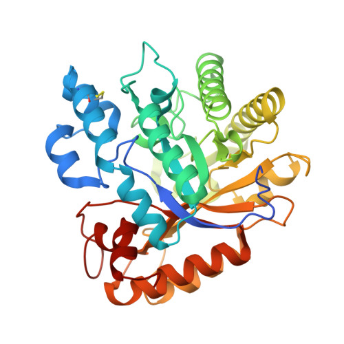
    Rutinosidases (α-l-rhamnosyl-β-d-glucosidases) catalyze the cleavage of the glycosidic bond between the aglycone and the disaccharide rutinose (α-l-rhamnopyranosyl-(1→6)-β-d-glucopyranose) of specific flavonoid glycosides such as rutin (quercetin 3-O-rutinoside). Microbial rutinosidases are part of the rutin catabolic pathway, enabling the microorganism to utilize rutin and related plant phenolic glycosides. Here, we report the first three-dimensional structure of a rutinosidase determined at 1.27-Å resolution. The rutinosidase from Aspergillus niger K2 (AnRut), a member of glycoside hydrolase family GH-5, subfamily 23, was heterologously produced in Pichia pastoris. The X-ray structure of AnRut is represented by a distorted (β/α) 8 barrel fold with its closest structural homologue being an exo-β-(1,3)-glucanase from Candida albicans (CaExg). The catalytic site is located in a deep pocket with a striking structural similarity to CaExg. However, the entrance to the active site of AnRut has been found to be different from that of CaExg - a mostly unstructured section of ~ 40 residues present in CaExg is missing in AnRut, whereas an additional loop of 13 amino acids partially covers the active site of AnRut. NMR analysis of reaction products provided clear evidence for a retaining reaction mechanism of AnRut. Unexpectedly, quercetin 3-O-glucoside was found to be a better substrate than rutin, and thus, AnRut cannot be considered a typical diglycosidase. Mutational analysis of conserved active site residues in combination with in silico modeling allowed identification of essential interactions for enzyme activity and helped to reveal further details of substrate binding. The protein sequence of AnRut has been revised. DATABASES: The nucleotide sequence of the rutinosidase-encoding gene is available in the GenBank database under the accession number MN393234. Structural data are available in the PDB database under the accession number 6I1A. ENZYME: α-l-Rhamnosyl-β-d-glucosidase (EC 3.2.1.168).


  • Organizational Affiliation

    Institute of Organic Chemistry and Biochemistry of the Czech Academy of Sciences, Prague, Czech Republic.


Macromolecules
Find similar proteins by:  (by identity cutoff)  |  3D Structure
Entity ID: 1
MoleculeChains Sequence LengthOrganismDetailsImage
rutinosidase
A, B
370Aspergillus nigerMutation(s): 0 
Gene Names: ATCC64974_105530
UniProt
Find proteins for G3YFQ1 (Aspergillus niger (strain ATCC 1015 / CBS 113.46 / FGSC A1144 / LSHB Ac4 / NCTC 3858a / NRRL 328 / USDA 3528.7))
Explore G3YFQ1 
Go to UniProtKB:  G3YFQ1
Entity Groups  
Sequence Clusters30% Identity50% Identity70% Identity90% Identity95% Identity100% Identity
UniProt GroupG3YFQ1
Sequence Annotations
Expand
  • Reference Sequence
Small Molecules
Ligands 2 Unique
IDChains Name / Formula / InChI Key2D Diagram3D Interactions
NAG
Query on NAG

Download Ideal Coordinates CCD File 
AA [auth B]
BA [auth B]
C [auth A]
D [auth A]
E [auth A]
AA [auth B],
BA [auth B],
C [auth A],
D [auth A],
E [auth A],
F [auth A],
G [auth A],
X [auth B],
Y [auth B],
Z [auth B]
2-acetamido-2-deoxy-beta-D-glucopyranose
C8 H15 N O6
OVRNDRQMDRJTHS-FMDGEEDCSA-N
EDO
Query on EDO

Download Ideal Coordinates CCD File 
CA [auth B]
DA [auth B]
EA [auth B]
FA [auth B]
GA [auth B]
CA [auth B],
DA [auth B],
EA [auth B],
FA [auth B],
GA [auth B],
H [auth A],
HA [auth B],
I [auth A],
IA [auth B],
J [auth A],
JA [auth B],
K [auth A],
KA [auth B],
L [auth A],
LA [auth B],
M [auth A],
MA [auth B],
N [auth A],
NA [auth B],
O [auth A],
P [auth A],
Q [auth A],
R [auth A],
S [auth A],
T [auth A],
U [auth A],
V [auth A],
W [auth A]
1,2-ETHANEDIOL
C2 H6 O2
LYCAIKOWRPUZTN-UHFFFAOYSA-N
Experimental Data & Validation

Experimental Data

  • Method: X-RAY DIFFRACTION
  • Resolution: 1.27 Å
  • R-Value Free: 0.178 
  • R-Value Work: 0.159 
  • R-Value Observed: 0.159 
  • Space Group: P 21 21 21
Unit Cell:
Length ( Å )Angle ( ˚ )
a = 53.068α = 90
b = 97.359β = 90
c = 139.259γ = 90
Software Package:
Software NamePurpose
REFMACrefinement
XDSdata reduction
XSCALEdata scaling
HKL2Mapphasing

Structure Validation

View Full Validation Report


Ligand Structure Quality Assessment 


Entry History & Funding Information

Deposition Data


Funding OrganizationLocationGrant Number
Czech Science FoundationCzech Republic18-00150S

Revision History  (Full details and data files)

  • Version 1.0: 2020-01-29
    Type: Initial release
  • Version 1.1: 2020-07-29
    Type: Remediation
    Reason: Carbohydrate remediation
    Changes: Data collection, Derived calculations, Refinement description, Structure summary
  • Version 1.2: 2020-08-12
    Changes: Database references, Structure summary