6HY0

Atomic models of P1, P4 C-terminal fragment and P8 fitted in the bacteriophage phi6 nucleocapsid reconstructed with icosahedral symmetry


Experimental Data Snapshot

  • Method: ELECTRON MICROSCOPY
  • Resolution: 3.50 Å
  • Aggregation State: PARTICLE 
  • Reconstruction Method: SINGLE PARTICLE 

wwPDB Validation   3D Report Full Report


This is version 1.2 of the entry. See complete history


Literature

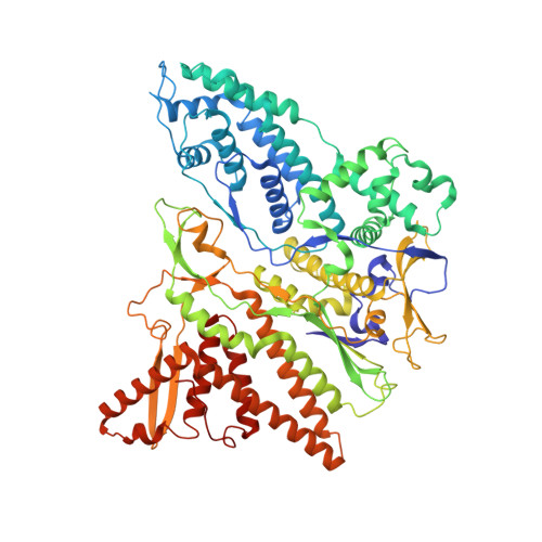
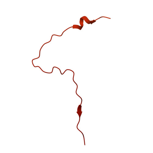
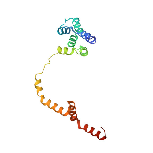
Multiple liquid crystalline geometries of highly compacted nucleic acid in a dsRNA virus.

Ilca, S.L.Sun, X.El Omari, K.Kotecha, A.de Haas, F.DiMaio, F.Grimes, J.M.Stuart, D.I.Poranen, M.M.Huiskonen, J.T.

(2019) Nature 570: 252-256

  • DOI: https://doi.org/10.1038/s41586-019-1229-9
  • Primary Citation of Related Structures:  
    6HY0

  • PubMed Abstract: 

    Characterizing the genome of mature virions is pivotal to understanding the highly dynamic processes of virus assembly and infection. Owing to the different cellular fates of DNA and RNA, the life cycles of double-stranded (ds)DNA and dsRNA viruses are dissimilar. In terms of nucleic acid packing, dsDNA viruses, which lack genome segmentation and intra-capsid transcriptional machinery, predominantly display single-spooled genome organizations 1-8 . Because the release of dsRNA into the cytoplasm triggers host defence mechanisms 9 , dsRNA viruses retain their genomes within a core particle that contains the enzymes required for RNA replication and transcription 10-12 . The genomes of dsRNA viruses vary greatly in the degree of segmentation. In members of the Reoviridae family, genomes consist of 10-12 segments and exhibit a non-spooled arrangement mediated by RNA-dependent RNA polymerases 11-14 . However, whether this arrangement is a general feature of dsRNA viruses remains unknown. Here, using cryo-electron microscopy to resolve the dsRNA genome structure of the tri-segmented bacteriophage ɸ6 of the Cystoviridae family, we show that dsRNA viruses can adopt a dsDNA-like single-spooled genome organization. We find that in this group of viruses, RNA-dependent RNA polymerases do not direct genome ordering, and the dsRNA can adopt multiple conformations. We build a model that encompasses 90% of the genome, and use this to quantify variation in the packing density and to characterize the different liquid crystalline geometries that are exhibited by the tightly compacted nucleic acid. Our results demonstrate that the canonical model for the packing of dsDNA can be extended to dsRNA viruses.


  • Organizational Affiliation

    Division of Structural Biology, Wellcome Centre for Human Genetics, University of Oxford, Oxford, UK.


Macromolecules
Find similar proteins by:  (by identity cutoff)  |  3D Structure
Entity ID: 1
MoleculeChains Sequence LengthOrganismDetailsImage
Major inner protein P1
A, B
769Cystovirus phi6Mutation(s): 0 
UniProt
Find proteins for P11126 (Pseudomonas phage phi6)
Explore P11126 
Go to UniProtKB:  P11126
Entity Groups  
Sequence Clusters30% Identity50% Identity70% Identity90% Identity95% Identity100% Identity
UniProt GroupP11126
Sequence Annotations
Expand
  • Reference Sequence
Find similar proteins by:  (by identity cutoff)  |  3D Structure
Entity ID: 2
MoleculeChains Sequence LengthOrganismDetailsImage
Packaging Enzyme P4332Cystovirus phi6Mutation(s): 0 
UniProt
Find proteins for P11125 (Pseudomonas phage phi6)
Explore P11125 
Go to UniProtKB:  P11125
Entity Groups  
Sequence Clusters30% Identity50% Identity70% Identity90% Identity95% Identity100% Identity
UniProt GroupP11125
Sequence Annotations
Expand
  • Reference Sequence
Find similar proteins by:  (by identity cutoff)  |  3D Structure
Entity ID: 3
MoleculeChains Sequence LengthOrganismDetailsImage
Major Outer Capsid Protein P8
D, E, F, G, H
D, E, F, G, H, I, J, K, L, M
149Cystovirus phi6Mutation(s): 0 
UniProt
Find proteins for P07579 (Pseudomonas phage phi6)
Explore P07579 
Go to UniProtKB:  P07579
Entity Groups  
Sequence Clusters30% Identity50% Identity70% Identity90% Identity95% Identity100% Identity
UniProt GroupP07579
Sequence Annotations
Expand
  • Reference Sequence
Experimental Data & Validation

Experimental Data

  • Method: ELECTRON MICROSCOPY
  • Resolution: 3.50 Å
  • Aggregation State: PARTICLE 
  • Reconstruction Method: SINGLE PARTICLE 
EM Software:
TaskSoftware PackageVersion
RECONSTRUCTIONRELION2.0.3
MODEL REFINEMENTREFMAC5

Structure Validation

View Full Validation Report


Entry History 

Deposition Data

Revision History  (Full details and data files)

  • Version 1.0: 2019-06-12
    Type: Initial release
  • Version 1.1: 2019-06-26
    Changes: Data collection, Database references, Derived calculations
  • Version 1.2: 2019-12-18
    Changes: Other