6HTM

X-ray structure of the tryptophan lyase NosL in complex with bound tryptamin


Experimental Data Snapshot

  • Method: X-RAY DIFFRACTION
  • Resolution: 1.70 Å
  • R-Value Free: 0.189 
  • R-Value Work: 0.162 
  • R-Value Observed: 0.164 

wwPDB Validation   3D Report Full Report


Ligand Structure Quality Assessment 


This is version 1.3 of the entry. See complete history


Literature

Radical S-Adenosyl-l-methionine Tryptophan Lyase (NosL): How the Protein Controls the Carboxyl Radical •CO2-Migration.

Amara, P.Mouesca, J.M.Bella, M.Martin, L.Saragaglia, C.Gambarelli, S.Nicolet, Y.

(2018) J Am Chem Soc 140: 16661-16668

  • DOI: https://doi.org/10.1021/jacs.8b09142
  • Primary Citation of Related Structures:  
    6HTK, 6HTM, 6HTO

  • PubMed Abstract: 

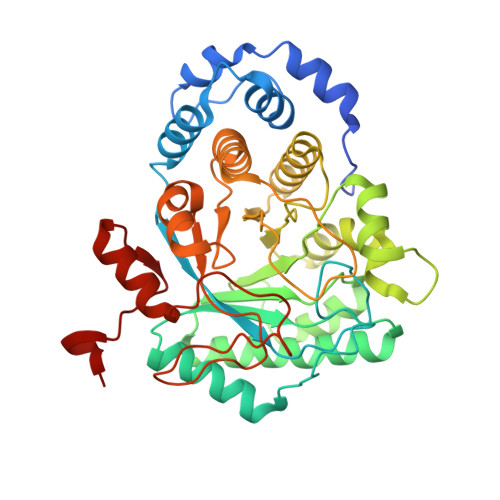
    The radical S-adenosyl-l-methionine tryptophan lyase uses radical-based chemistry to convert l-tryptophan into 3-methyl-2-indolic acid, a fragment in the biosynthesis of the thiopeptide antibiotic nosiheptide. This complex reaction involves several successive steps corresponding to (i) the activation by a specific hydrogen-atom abstraction, (ii) an unprecedented •CO 2 - radical migration, (iii) a cyanide fragment release, and (iv) the termination of the radical-based reaction. In vitro study of this reaction is made more difficult because the enzyme produces a significant amount of a shunt product instead of the natural product. Here, using a combination of X-ray crystallography, electron paramagnetic resonance spectroscopy, and quantum and hybrid quantum mechanical/molecular mechanical calculations, we have deciphered the fine mechanism of the key •CO 2 - radical migration, highlighting how the preorganized active site of the protein tightly controls this reaction.


  • Organizational Affiliation

    Univ. Grenoble Alpes, CEA, CNRS, IBS , Metalloproteins Unit , F-38000 Grenoble , France.


Macromolecules
Find similar proteins by:  (by identity cutoff)  |  3D Structure
Entity ID: 1
MoleculeChains Sequence LengthOrganismDetailsImage
3-methyl-2-indolic acid synthase
A, B
400Streptomyces actuosusMutation(s): 0 
Gene Names: nosLnocLDMT42_23170
UniProt
Find proteins for C6FX51 (Streptomyces actuosus)
Explore C6FX51 
Go to UniProtKB:  C6FX51
Entity Groups  
Sequence Clusters30% Identity50% Identity70% Identity90% Identity95% Identity100% Identity
UniProt GroupC6FX51
Sequence Annotations
Expand
  • Reference Sequence
Small Molecules
Ligands 8 Unique
IDChains Name / Formula / InChI Key2D Diagram3D Interactions
SF4
Query on SF4

Download Ideal Coordinates CCD File 
D [auth A],
N [auth B]
IRON/SULFUR CLUSTER
Fe4 S4
LJBDFODJNLIPKO-UHFFFAOYSA-N
5AD
Query on 5AD

Download Ideal Coordinates CCD File 
F [auth A],
Q [auth B]
5'-DEOXYADENOSINE
C10 H13 N5 O3
XGYIMTFOTBMPFP-KQYNXXCUSA-N
TSS
Query on TSS

Download Ideal Coordinates CCD File 
C [auth A],
O [auth B]
2-(1H-INDOL-3-YL)ETHANAMINE
C10 H12 N2
APJYDQYYACXCRM-UHFFFAOYSA-N
MET
Query on MET

Download Ideal Coordinates CCD File 
E [auth A],
P [auth B]
METHIONINE
C5 H11 N O2 S
FFEARJCKVFRZRR-BYPYZUCNSA-N
GOL
Query on GOL

Download Ideal Coordinates CCD File 
G [auth A]
H [auth A]
R [auth B]
S [auth B]
T [auth B]
G [auth A],
H [auth A],
R [auth B],
S [auth B],
T [auth B],
U [auth B]
GLYCEROL
C3 H8 O3
PEDCQBHIVMGVHV-UHFFFAOYSA-N
BR
Query on BR

Download Ideal Coordinates CCD File 
I [auth A]
J [auth A]
K [auth A]
V [auth B]
W [auth B]
I [auth A],
J [auth A],
K [auth A],
V [auth B],
W [auth B],
X [auth B]
BROMIDE ION
Br
CPELXLSAUQHCOX-UHFFFAOYSA-M
ZN
Query on ZN

Download Ideal Coordinates CCD File 
BA [auth B]ZINC ION
Zn
PTFCDOFLOPIGGS-UHFFFAOYSA-N
CL
Query on CL

Download Ideal Coordinates CCD File 
AA [auth B],
L [auth A],
M [auth A],
Y [auth B],
Z [auth B]
CHLORIDE ION
Cl
VEXZGXHMUGYJMC-UHFFFAOYSA-M
Experimental Data & Validation

Experimental Data

  • Method: X-RAY DIFFRACTION
  • Resolution: 1.70 Å
  • R-Value Free: 0.189 
  • R-Value Work: 0.162 
  • R-Value Observed: 0.164 
  • Space Group: P 1 2 1
Unit Cell:
Length ( Å )Angle ( ˚ )
a = 94.53α = 90
b = 47.14β = 108.9
c = 113.9γ = 90
Software Package:
Software NamePurpose
PHENIXrefinement
XDSdata reduction
XSCALEdata scaling
PHENIXphasing

Structure Validation

View Full Validation Report


Ligand Structure Quality Assessment 


Entry History & Funding Information

Deposition Data


Funding OrganizationLocationGrant Number
French National Research AgencyFranceANR-16-CE29-0019

Revision History  (Full details and data files)

  • Version 1.0: 2018-11-21
    Type: Initial release
  • Version 1.1: 2018-11-28
    Changes: Data collection, Database references
  • Version 1.2: 2019-04-24
    Changes: Data collection, Database references
  • Version 1.3: 2024-01-24
    Changes: Data collection, Database references, Derived calculations, Refinement description