6H8T

Crystal structure of Papain modify by achiral Ru(II)complex


Experimental Data Snapshot

  • Method: X-RAY DIFFRACTION
  • Resolution: 2.10 Å
  • R-Value Free: 0.249 
  • R-Value Work: 0.192 
  • R-Value Observed: 0.195 

wwPDB Validation   3D Report Full Report


Ligand Structure Quality Assessment 


This is version 1.2 of the entry. See complete history

Re-refinement Note

This entry reflects an alternative modeling of the original data in: 4KP9


Literature

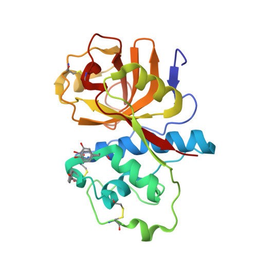
Crystallographic evidence for unexpected selective tyrosine hydroxylations in an aerated achiral Ru-papain conjugate.

Cherrier, M.V.Amara, P.Talbi, B.Salmain, M.Fontecilla-Camps, J.C.

(2018) Metallomics 10: 1452-1459

  • DOI: https://doi.org/10.1039/c8mt00160j
  • Primary Citation of Related Structures:  
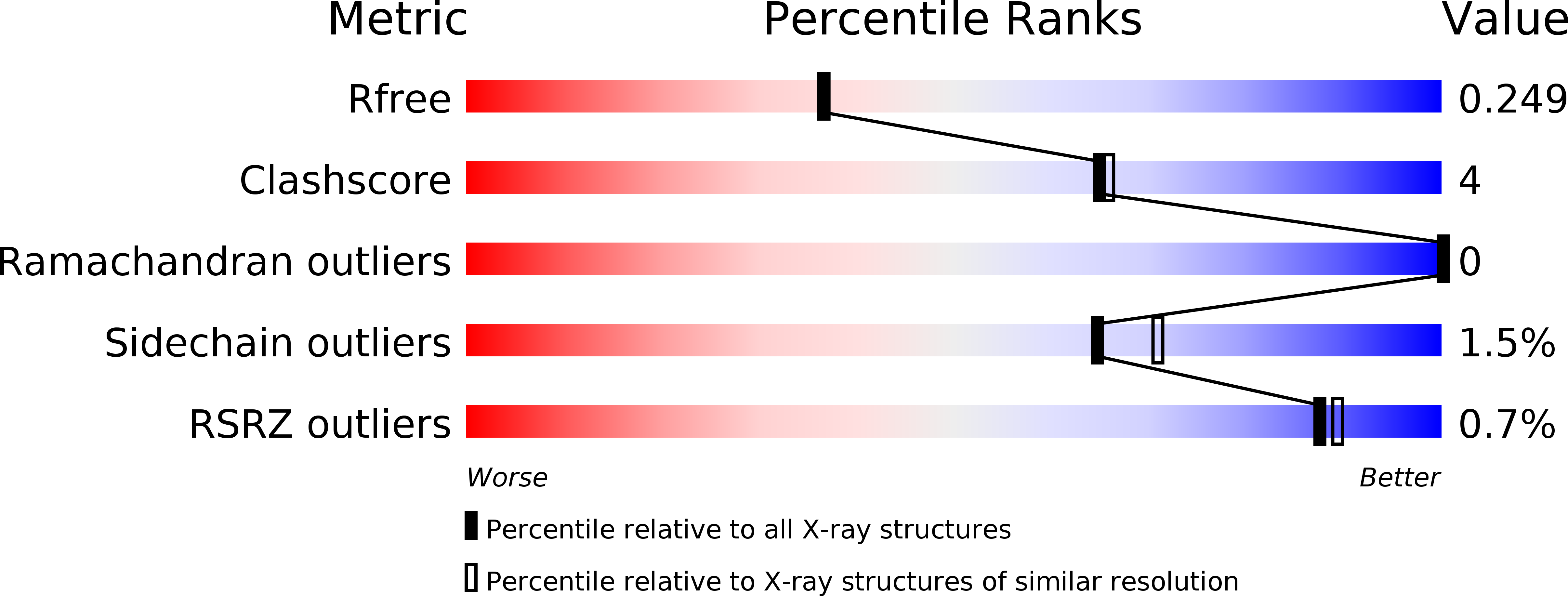
    6H8T

  • PubMed Abstract: 

    The X-ray structure of an aerated achiral Ru-papain conjugate has revealed the hydroxylation of two tyrosine residues found near the ruthenium ion. The most likely mechanism involves a ruthenium-bound superoxide as the reactive species responsible for the first hydroxylation and the resulting high valent Ru(iv)[double bond, length as m-dash]O species for the second one.


  • Organizational Affiliation

    Univ. Grenoble Alpes, CEA, CNRS, IBS, Metalloproteins, F-38000 Grenoble, France. juan.fontecilla@ibs.fr.


Macromolecules
Find similar proteins by:  (by identity cutoff)  |  3D Structure
Entity ID: 1
MoleculeChains Sequence LengthOrganismDetailsImage
PapainA,
B [auth J]
212Carica papayaMutation(s): 0 
EC: 3.4.22.2
UniProt
Find proteins for P00784 (Carica papaya)
Explore P00784 
Go to UniProtKB:  P00784
Entity Groups  
Sequence Clusters30% Identity50% Identity70% Identity90% Identity95% Identity100% Identity
UniProt GroupP00784
Sequence Annotations
Expand
  • Reference Sequence
Small Molecules
Ligands 6 Unique
IDChains Name / Formula / InChI Key2D Diagram3D Interactions
YXX
Query on YXX

Download Ideal Coordinates CCD File 
H [auth J]chloro(1-{5-[di(pyridin-2-yl-kappaN)amino]pentyl}pyrrolidine-2,5-dione)ruthenium
C19 H22 Cl N4 O2 Ru
LYRJXCXFYIQAEL-UHFFFAOYSA-M
YXZ
Query on YXZ

Download Ideal Coordinates CCD File 
G [auth A]chloro[N,N-di(pyridin-2-yl-kappaN)pentane-1,5-diamine]ruthenium
C15 H20 Cl N4 Ru
IIQHAXSUPAXJMA-UHFFFAOYSA-M
SO4
Query on SO4

Download Ideal Coordinates CCD File 
I [auth J]SULFATE ION
O4 S
QAOWNCQODCNURD-UHFFFAOYSA-L
ACT
Query on ACT

Download Ideal Coordinates CCD File 
C [auth A]
E [auth A]
F [auth A]
J
K [auth J]
C [auth A],
E [auth A],
F [auth A],
J,
K [auth J],
M [auth J],
N [auth J],
O [auth J],
P [auth J]
ACETATE ION
C2 H3 O2
QTBSBXVTEAMEQO-UHFFFAOYSA-M
CL
Query on CL

Download Ideal Coordinates CCD File 
D [auth A]CHLORIDE ION
Cl
VEXZGXHMUGYJMC-UHFFFAOYSA-M
NA
Query on NA

Download Ideal Coordinates CCD File 
L [auth J]SODIUM ION
Na
FKNQFGJONOIPTF-UHFFFAOYSA-N
Modified Residues  2 Unique
IDChains TypeFormula2D DiagramParent
OMX
Query on OMX
A,
B [auth J]
L-PEPTIDE LINKINGC9 H11 N O4TYR
TXY
Query on TXY
A,
B [auth J]
L-PEPTIDE LINKINGC9 H13 N O4TYR
Experimental Data & Validation

Experimental Data

  • Method: X-RAY DIFFRACTION
  • Resolution: 2.10 Å
  • R-Value Free: 0.249 
  • R-Value Work: 0.192 
  • R-Value Observed: 0.195 
  • Space Group: P 21 21 21
Unit Cell:
Length ( Å )Angle ( ˚ )
a = 61.03α = 90
b = 74.85β = 90
c = 86.25γ = 90
Software Package:
Software NamePurpose
PHENIXrefinement
XDSdata reduction
XSCALEdata scaling
PHASERphasing

Structure Validation

View Full Validation Report


Ligand Structure Quality Assessment 


Entry History & Funding Information

Deposition Data


Funding OrganizationLocationGrant Number
French National Research AgencyFranceARTZYME

Revision History  (Full details and data files)

  • Version 1.0: 2018-09-12
    Type: Initial release
  • Version 1.1: 2018-10-24
    Changes: Data collection, Database references
  • Version 1.2: 2024-01-17
    Changes: Data collection, Database references, Refinement description