6GVJ

Human Mps1 kinase domain with ordered activation loop


Experimental Data Snapshot

  • Method: X-RAY DIFFRACTION
  • Resolution: 2.41 Å
  • R-Value Free: 0.233 
  • R-Value Work: 0.201 
  • R-Value Observed: 0.203 

wwPDB Validation   3D Report Full Report


This is version 1.2 of the entry. See complete history


Literature

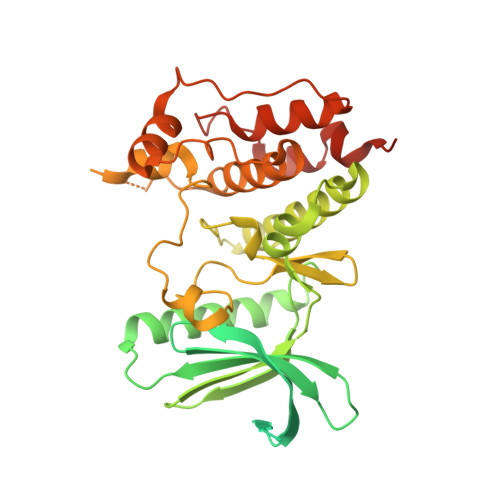
A crystal structure of the human protein kinase Mps1 reveals an ordered conformation of the activation loop.

Roorda, J.C.Joosten, R.P.Perrakis, A.Hiruma, Y.

(2019) Proteins 87: 348-352

  • DOI: https://doi.org/10.1002/prot.25651
  • Primary Citation of Related Structures:  
    6GVJ

  • PubMed Abstract: 

    Monopolar spindle 1 (Mps1) is a dual-specificity protein kinase, orchestrating faithful chromosome segregation during mitosis. All reported structures of the Mps1 kinase adopt the hallmarks of an inactive conformation, which includes a mostly disordered activation loop. Here, we present a 2.4 Å resolution crystal structure of an "extended" version of the Mps1 kinase domain, which shows an ordered activation loop. However, the other structural characteristics of an active kinase are not present. Our structure shows that the Mps1 activation loop can fit to the ATP binding pocket and interferes with ATP, but less so with inhibitors binding, partly explain the potency of various Mps1 inhibitors.


  • Organizational Affiliation

    Division of Biochemistry, Netherlands Cancer Institute, Amsterdam, The Netherlands.


Macromolecules
Find similar proteins by:  (by identity cutoff)  |  3D Structure
Entity ID: 1
MoleculeChains Sequence LengthOrganismDetailsImage
Dual specificity protein kinase TTK429Homo sapiensMutation(s): 0 
Gene Names: TTKMPS1MPS1L1
EC: 2.7.12.1
UniProt & NIH Common Fund Data Resources
Find proteins for P33981 (Homo sapiens)
Explore P33981 
Go to UniProtKB:  P33981
PHAROS:  P33981
GTEx:  ENSG00000112742 
Entity Groups  
Sequence Clusters30% Identity50% Identity70% Identity90% Identity95% Identity100% Identity
UniProt GroupP33981
Sequence Annotations
Expand
  • Reference Sequence
Experimental Data & Validation

Experimental Data

  • Method: X-RAY DIFFRACTION
  • Resolution: 2.41 Å
  • R-Value Free: 0.233 
  • R-Value Work: 0.201 
  • R-Value Observed: 0.203 
  • Space Group: I 2 2 2
Unit Cell:
Length ( Å )Angle ( ˚ )
a = 70.825α = 90
b = 102.613β = 90
c = 110.973γ = 90
Software Package:
Software NamePurpose
REFMACrefinement
Aimlessdata scaling
MOLREPphasing
PDB_EXTRACTdata extraction
xia2data reduction

Structure Validation

View Full Validation Report


Entry History & Funding Information

Deposition Data


Funding OrganizationLocationGrant Number
Netherlands Organisation for Scientific ResearchNetherlands722.015.008
Netherlands Organisation for Scientific ResearchNetherlands723.013.003

Revision History  (Full details and data files)

  • Version 1.0: 2019-01-09
    Type: Initial release
  • Version 1.1: 2019-03-20
    Changes: Data collection, Database references
  • Version 1.2: 2024-01-17
    Changes: Data collection, Database references, Refinement description