6GDY

Crystal structure of 2OG oxygenase JMJD6 (aa 1-343) in complex with Fe(II) and 2OG


Experimental Data Snapshot

  • Method: X-RAY DIFFRACTION
  • Resolution: 2.04 Å
  • R-Value Free: 0.216 
  • R-Value Work: 0.179 
  • R-Value Observed: 0.181 

wwPDB Validation   3D Report Full Report


This is version 1.1 of the entry. See complete history


Literature

Biochemical and structural investigations clarify the substrate selectivity of the 2-oxoglutarate oxygenase JMJD6.

Islam, M.S.McDonough, M.A.Chowdhury, R.Gault, J.Khan, A.Pires, E.Schofield, C.J.

(2019) J Biol Chem 294: 11637-11652

  • DOI: https://doi.org/10.1074/jbc.RA119.008693
  • Primary Citation of Related Structures:  
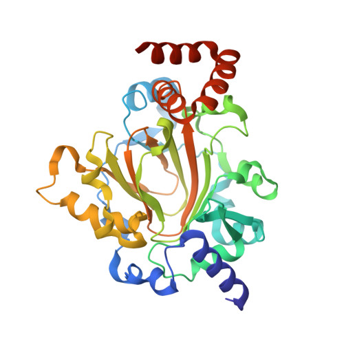
    6GDY

  • PubMed Abstract: 

    JmjC domain-containing protein 6 (JMJD6) is a 2-oxoglutarate (2OG)-dependent oxygenase linked to various cellular processes, including splicing regulation, histone modification, transcriptional pause release, hypoxia sensing, and cancer. JMJD6 is reported to catalyze hydroxylation of lysine residue(s) of histones, the tumor-suppressor protein p53, and splicing regulatory proteins, including u2 small nuclear ribonucleoprotein auxiliary factor 65-kDa subunit (U2AF65). JMJD6 is also reported to catalyze N -demethylation of N -methylated (both mono- and di-methylated) arginine residues of histones and other proteins, including HSP70 (heat-shock protein 70), estrogen receptor α, and RNA helicase A. Here, we report MS- and NMR-based kinetic assays employing purified JMJD6 and multiple substrate fragment sequences, the results of which support the assignment of purified JMJD6 as a lysyl hydroxylase. By contrast, we did not observe N -methyl arginyl N -demethylation with purified JMJD6. Biophysical analyses, including crystallographic analyses of JMJD6 Δ344-403 in complex with iron and 2OG, supported its assignment as a lysyl hydroxylase rather than an N -methyl arginyl-demethylase. The screening results supported some, but not all, of the assigned JMJD6 substrates and identified other potential JMJD6 substrates. We envision these results will be useful in cellular and biological work on the substrates and functions of JMJD6 and in the development of selective inhibitors of human 2OG oxygenases.


  • Organizational Affiliation

    Department of Chemistry, University of Oxford, 12 Mansfield Road, Oxford OX1 3TA, United Kingdom.


Macromolecules
Find similar proteins by:  (by identity cutoff)  |  3D Structure
Entity ID: 1
MoleculeChains Sequence LengthOrganismDetailsImage
Bifunctional arginine demethylase and lysyl-hydroxylase JMJD6
A, B
343Homo sapiensMutation(s): 0 
Gene Names: JMJD6KIAA0585PTDSR
EC: 1.14.11
UniProt & NIH Common Fund Data Resources
Find proteins for Q6NYC1 (Homo sapiens)
Explore Q6NYC1 
Go to UniProtKB:  Q6NYC1
PHAROS:  Q6NYC1
GTEx:  ENSG00000070495 
Entity Groups  
Sequence Clusters30% Identity50% Identity70% Identity90% Identity95% Identity100% Identity
UniProt GroupQ6NYC1
Sequence Annotations
Expand
  • Reference Sequence
Small Molecules
Ligands 6 Unique
IDChains Name / Formula / InChI Key2D Diagram3D Interactions
EPE
Query on EPE

Download Ideal Coordinates CCD File 
K [auth A],
L [auth A]
4-(2-HYDROXYETHYL)-1-PIPERAZINE ETHANESULFONIC ACID
C8 H18 N2 O4 S
JKMHFZQWWAIEOD-UHFFFAOYSA-N
AKG
Query on AKG

Download Ideal Coordinates CCD File 
D [auth A],
O [auth B]
2-OXOGLUTARIC ACID
C5 H6 O5
KPGXRSRHYNQIFN-UHFFFAOYSA-N
PO4
Query on PO4

Download Ideal Coordinates CCD File 
F [auth A]
G [auth A]
H [auth A]
I [auth A]
J [auth A]
F [auth A],
G [auth A],
H [auth A],
I [auth A],
J [auth A],
P [auth B],
Q [auth B],
R [auth B],
S [auth B],
T [auth B]
PHOSPHATE ION
O4 P
NBIIXXVUZAFLBC-UHFFFAOYSA-K
GOL
Query on GOL

Download Ideal Coordinates CCD File 
M [auth A],
U [auth B],
V [auth B]
GLYCEROL
C3 H8 O3
PEDCQBHIVMGVHV-UHFFFAOYSA-N
FE
Query on FE

Download Ideal Coordinates CCD File 
C [auth A],
N [auth B]
FE (III) ION
Fe
VTLYFUHAOXGGBS-UHFFFAOYSA-N
NA
Query on NA

Download Ideal Coordinates CCD File 
E [auth A]SODIUM ION
Na
FKNQFGJONOIPTF-UHFFFAOYSA-N
Binding Affinity Annotations 
IDSourceBinding Affinity
AKG Binding MOAD:  6GDY Kd: 3500 (nM) from 1 assay(s)
Experimental Data & Validation

Experimental Data

  • Method: X-RAY DIFFRACTION
  • Resolution: 2.04 Å
  • R-Value Free: 0.216 
  • R-Value Work: 0.179 
  • R-Value Observed: 0.181 
  • Space Group: P 1 21 1
Unit Cell:
Length ( Å )Angle ( ˚ )
a = 47.15α = 90
b = 96.99β = 95.84
c = 93.76γ = 90
Software Package:
Software NamePurpose
PHENIXrefinement
XDSdata reduction
SCALAdata scaling
SHARPphasing

Structure Validation

View Full Validation Report


Entry History & Funding Information

Deposition Data


Funding OrganizationLocationGrant Number
United Kingdom--

Revision History  (Full details and data files)

  • Version 1.0: 2019-04-03
    Type: Initial release
  • Version 1.1: 2019-10-16
    Changes: Data collection, Database references