6FKQ

THE CRYSTAL STRUCTURE OF A FRAGMENT OF NETRIN-1 IN COMPLEX WITH A FRAGMENT OF DRAXIN


Experimental Data Snapshot

  • Method: X-RAY DIFFRACTION
  • Resolution: 3.07 Å
  • R-Value Free: 0.271 
  • R-Value Work: 0.235 
  • R-Value Observed: 0.237 

wwPDB Validation   3D Report Full Report


This is version 2.1 of the entry. See complete history


Literature

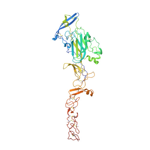
Structural Basis for Draxin-Modulated Axon Guidance and Fasciculation by Netrin-1 through DCC.

Liu, Y.Bhowmick, T.Liu, Y.Gao, X.Mertens, H.D.T.Svergun, D.I.Xiao, J.Zhang, Y.Wang, J.H.Meijers, R.

(2018) Neuron 97: 1261-1267.e4

  • DOI: https://doi.org/10.1016/j.neuron.2018.02.010
  • Primary Citation of Related Structures:  
    5Z5K, 6FKQ

  • PubMed Abstract: 

    Axon guidance involves the spatiotemporal interplay between guidance cues and membrane-bound cell-surface receptors, present on the growth cone of the axon. Netrin-1 is a prototypical guidance cue that binds to deleted in colorectal cancer (DCC), and it has been proposed that the guidance cue Draxin modulates this interaction. Here, we present structural snapshots of Draxin/DCC and Draxin/Netrin-1 complexes, revealing a triangular relationship that affects Netrin-mediated haptotaxis and fasciculation. Draxin interacts with DCC through the N-terminal four immunoglobulin domains, and Netrin-1 through the EGF-3 domain, in the same region where DCC binds. Netrin-1 and DCC bind to adjacent sites on Draxin, which appears to capture Netrin-1 and tether it to the DCC receptor. We propose the conformational flexibility of the single-pass membrane receptor DCC is used to promote fasciculation and regulate axon guidance through concerted Netrin-1/Draxin binding. VIDEO ABSTRACT.


  • Organizational Affiliation

    State Key Laboratory of Membrane Biology, College of Life Sciences, Peking University, Beijing 100871, China.


Macromolecules
Find similar proteins by:  (by identity cutoff)  |  3D Structure
Entity ID: 1
MoleculeChains Sequence LengthOrganismDetailsImage
Netrin-1415Homo sapiensMutation(s): 0 
Gene Names: NTN1NTN1L
UniProt & NIH Common Fund Data Resources
Find proteins for O95631 (Homo sapiens)
Explore O95631 
Go to UniProtKB:  O95631
PHAROS:  O95631
GTEx:  ENSG00000065320 
Entity Groups  
Sequence Clusters30% Identity50% Identity70% Identity90% Identity95% Identity100% Identity
UniProt GroupO95631
Sequence Annotations
Expand
  • Reference Sequence

Find similar proteins by:  Sequence   |   3D Structure  

Entity ID: 2
MoleculeChains Sequence LengthOrganismDetailsImage
Draxin19Homo sapiensMutation(s): 0 
Gene Names: DRAXINC1orf187PSEC0258UNQ3119/PRO10268
UniProt & NIH Common Fund Data Resources
Find proteins for Q8NBI3 (Homo sapiens)
Explore Q8NBI3 
Go to UniProtKB:  Q8NBI3
PHAROS:  Q8NBI3
GTEx:  ENSG00000162490 
Entity Groups  
Sequence Clusters30% Identity50% Identity70% Identity90% Identity95% Identity100% Identity
UniProt GroupQ8NBI3
Sequence Annotations
Expand
  • Reference Sequence
Oligosaccharides

Help

Entity ID: 3
MoleculeChains Length2D Diagram Glycosylation3D Interactions
2-acetamido-2-deoxy-beta-D-glucopyranose-(1-4)-2-acetamido-2-deoxy-beta-D-glucopyranose
C, E
2N-Glycosylation
Glycosylation Resources
GlyTouCan:  G42666HT
GlyCosmos:  G42666HT
GlyGen:  G42666HT
Entity ID: 4
MoleculeChains Length2D Diagram Glycosylation3D Interactions
2-acetamido-2-deoxy-beta-D-glucopyranose-(1-4)-2-acetamido-2-deoxy-beta-D-glucopyranose-(1-4)-2-acetamido-2-deoxy-beta-D-glucopyranose
D
3N-Glycosylation
Glycosylation Resources
GlyTouCan:  G47362BJ
GlyCosmos:  G47362BJ
GlyGen:  G47362BJ
Small Molecules
Ligands 3 Unique
IDChains Name / Formula / InChI Key2D Diagram3D Interactions
SO4
Query on SO4

Download Ideal Coordinates CCD File 
G [auth A]
H [auth A]
I [auth A]
J [auth A]
K [auth A]
G [auth A],
H [auth A],
I [auth A],
J [auth A],
K [auth A],
L [auth A],
M [auth A],
N [auth A],
O [auth A],
P [auth A]
SULFATE ION
O4 S
QAOWNCQODCNURD-UHFFFAOYSA-L
GOL
Query on GOL

Download Ideal Coordinates CCD File 
Q [auth A]GLYCEROL
C3 H8 O3
PEDCQBHIVMGVHV-UHFFFAOYSA-N
CA
Query on CA

Download Ideal Coordinates CCD File 
F [auth A]CALCIUM ION
Ca
BHPQYMZQTOCNFJ-UHFFFAOYSA-N
Biologically Interesting Molecules (External Reference) 1 Unique
Experimental Data & Validation

Experimental Data

  • Method: X-RAY DIFFRACTION
  • Resolution: 3.07 Å
  • R-Value Free: 0.271 
  • R-Value Work: 0.235 
  • R-Value Observed: 0.237 
  • Space Group: P 64 2 2
Unit Cell:
Length ( Å )Angle ( ˚ )
a = 130.794α = 90
b = 130.794β = 90
c = 183.87γ = 120
Software Package:
Software NamePurpose
REFMACrefinement
MOSFLMdata reduction
Aimlessdata scaling
PHASERphasing

Structure Validation

View Full Validation Report


Entry History & Funding Information

Deposition Data


Funding OrganizationLocationGrant Number
Marie Curie CoFUNDGermany--

Revision History  (Full details and data files)

  • Version 1.0: 2018-03-21
    Type: Initial release
  • Version 1.1: 2018-04-04
    Changes: Data collection, Database references
  • Version 2.0: 2020-07-29
    Type: Remediation
    Reason: Carbohydrate remediation
    Changes: Advisory, Atomic model, Data collection, Derived calculations, Structure summary
  • Version 2.1: 2024-01-17
    Changes: Data collection, Database references, Derived calculations, Refinement description, Structure summary