6FDN

Rio2 structure


Experimental Data Snapshot

  • Method: X-RAY DIFFRACTION
  • Resolution: 2.90 Å
  • R-Value Free: 0.267 
  • R-Value Work: 0.226 
  • R-Value Observed: 0.228 

wwPDB Validation   3D Report Full Report


This is version 1.3 of the entry. See complete history


Literature

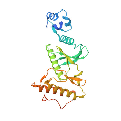
In vitro dimerization of human RIO2 kinase.

Maurice, F.Perebaskine, N.Thore, S.Fribourg, S.

(2019) RNA Biol 16: 1633-1642

  • DOI: https://doi.org/10.1080/15476286.2019.1653679
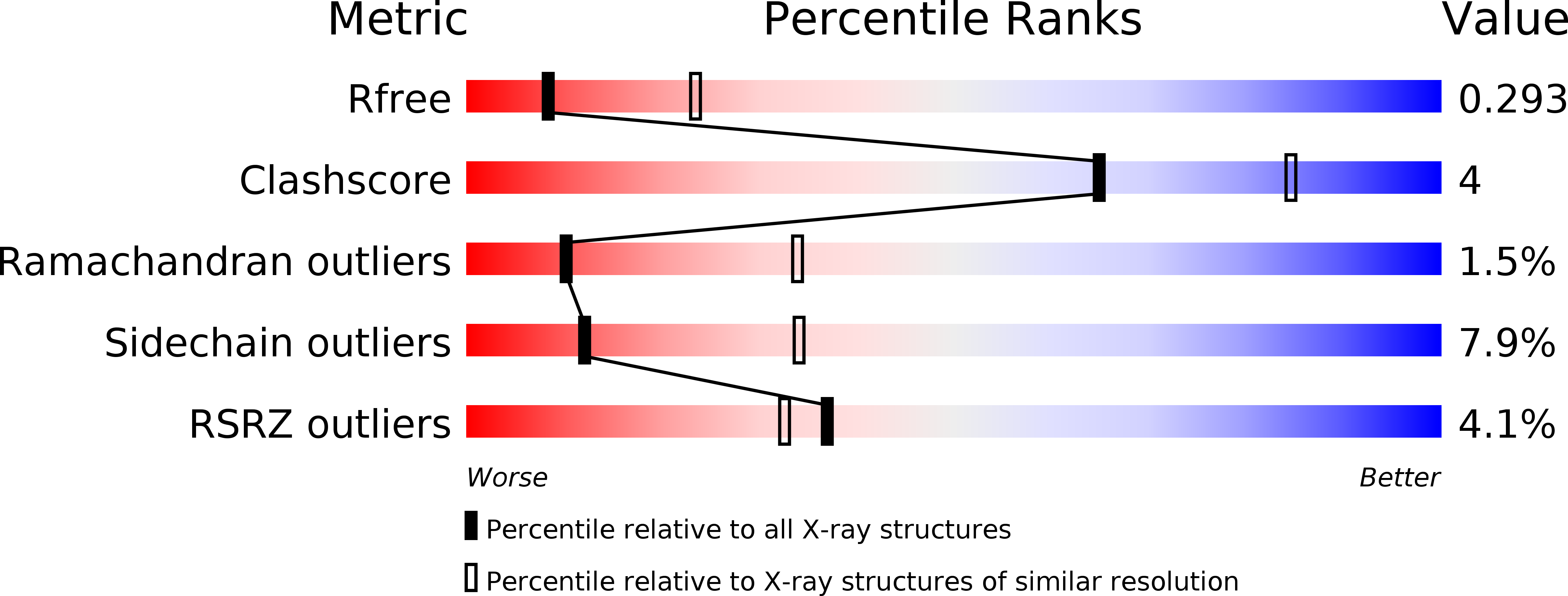
  • Primary Citation of Related Structures:  
    6FDM, 6FDN, 6FDO

  • PubMed Abstract: 

    RIO proteins form a conserved family of atypical protein kinases. RIO2 is a serine/threonine protein kinase/ATPase involved in pre-40S ribosomal maturation. Current crystal structures of archaeal and fungal Rio2 proteins report a monomeric form of the protein. Here, we describe three atomic structures of the human RIO2 kinase showing that it forms a homodimer in vitro . Upon self-association, each protomer ATP-binding pocket is partially remodelled and found in an apostate. The homodimerization is mediated by key residues previously shown to be responsible for ATP binding and catalysis. This unusual in vitro protein kinase dimer reveals an intricate mechanism where identical residues are involved in substrate binding and oligomeric state formation. We speculate that such an oligomeric state might be formed also in vivo and might function in maintaining the protein in an inactive state and could be employed during import.


  • Organizational Affiliation

    INSERM U1212, UMR CNRS 5320, Université de Bordeaux , Bordeaux , France.


Macromolecules
Find similar proteins by:  (by identity cutoff)  |  3D Structure
Entity ID: 1
MoleculeChains Sequence LengthOrganismDetailsImage
Serine/threonine-protein kinase RIO2
A, B
320Homo sapiensMutation(s): 0 
Gene Names: RIOK2
EC: 2.7.11.1
UniProt & NIH Common Fund Data Resources
Find proteins for Q9BVS4 (Homo sapiens)
Explore Q9BVS4 
Go to UniProtKB:  Q9BVS4
PHAROS:  Q9BVS4
GTEx:  ENSG00000058729 
Entity Groups  
Sequence Clusters30% Identity50% Identity70% Identity90% Identity95% Identity100% Identity
UniProt GroupQ9BVS4
Sequence Annotations
Expand
  • Reference Sequence
Experimental Data & Validation

Experimental Data

  • Method: X-RAY DIFFRACTION
  • Resolution: 2.90 Å
  • R-Value Free: 0.267 
  • R-Value Work: 0.226 
  • R-Value Observed: 0.228 
  • Space Group: P 21 21 21
Unit Cell:
Length ( Å )Angle ( ˚ )
a = 58.592α = 90
b = 86.405β = 90
c = 121.544γ = 90
Software Package:
Software NamePurpose
BUSTERrefinement
XDSdata reduction
XSCALEdata scaling

Structure Validation

View Full Validation Report


Entry History & Funding Information

Deposition Data

  • Released Date: 2019-01-30 
  • Deposition Author(s): Fribourg, S.

Funding OrganizationLocationGrant Number
French National Research AgencyFranceANR- RIBPRE40S

Revision History  (Full details and data files)

  • Version 1.0: 2019-01-30
    Type: Initial release
  • Version 1.1: 2019-08-28
    Changes: Data collection
  • Version 1.2: 2019-09-11
    Changes: Data collection, Database references
  • Version 1.3: 2019-10-09
    Changes: Data collection, Database references