6EWW

Structure of 14-3-3 zeta in complex with CaMKK2 14-3-3 binding motif


Experimental Data Snapshot

  • Method: X-RAY DIFFRACTION
  • Resolution: 2.68 Å
  • R-Value Free: 0.234 
  • R-Value Work: 0.208 
  • R-Value Observed: 0.209 

wwPDB Validation   3D Report Full Report


This is version 1.4 of the entry. See complete history


Literature

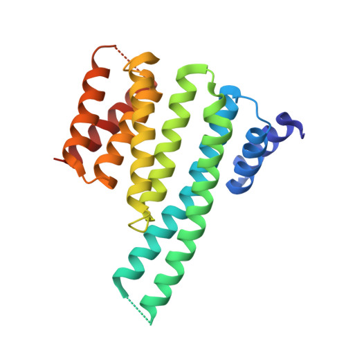
14-3-3 protein directly interacts with the kinase domain of calcium/calmodulin-dependent protein kinase kinase (CaMKK2).

Psenakova, K.Petrvalska, O.Kylarova, S.Lentini Santo, D.Kalabova, D.Herman, P.Obsilova, V.Obsil, T.

(2018) Biochim Biophys Acta 1862: 1612-1625

  • DOI: https://doi.org/10.1016/j.bbagen.2018.04.006
  • Primary Citation of Related Structures:  
    6EWW, 6FEL

  • PubMed Abstract: 

    Calcium/calmodulin-dependent protein kinase kinase 2 (CaMKK2) is a member of the Ca 2+ /calmodulin-dependent kinase (CaMK) family involved in adiposity regulation, glucose homeostasis and cancer. This upstream activator of CaMKI, CaMKIV and AMP-activated protein kinase is inhibited by phosphorylation, which also triggers an association with the scaffolding protein 14-3-3. However, the role of 14-3-3 in the regulation of CaMKK2 remains unknown. The interaction between phosphorylated CaMKK2 and the 14-3-3γ protein, as well as the architecture of their complex, were studied using enzyme activity measurements, small-angle x-ray scattering (SAXS), time-resolved fluorescence spectroscopy and protein crystallography. Our data suggest that the 14-3-3 protein binding does not inhibit the catalytic activity of phosphorylated CaMKK2 but rather slows down its dephosphorylation. Structural analysis indicated that the complex is flexible and that CaMKK2 is located outside the phosphopeptide-binding central channel of the 14-3-3γ dimer. Furthermore, 14-3-3γ appears to interact with and affect the structure of several regions of CaMKK2 outside the 14-3-3 binding motifs. In addition, the structural basis of interactions between 14-3-3 and the 14-3-3 binding motifs of CaMKK2 were elucidated by determining the crystal structures of phosphopeptides containing these motifs bound to 14-3-3. 14-3-3γ protein directly interacts with the kinase domain of CaMKK2 and the region containing the inhibitory phosphorylation site Thr 145 within the N-terminal extension. Our results suggested that CaMKK isoforms differ in their 14-3-3-mediated regulations and that the interaction between 14-3-3 protein and the N-terminal 14-3-3-binding motif of CaMKK2 might be stabilized by small-molecule compounds.


  • Organizational Affiliation

    Department of Physical and Macromolecular Chemistry, Faculty of Science, Charles University, Prague, Czech Republic; BioCeV - Institute of Physiology, The Czech Academy of Sciences, Vestec, Czech Republic.


Macromolecules
Find similar proteins by:  (by identity cutoff)  |  3D Structure
Entity ID: 1
MoleculeChains Sequence LengthOrganismDetailsImage
14-3-3 protein zeta/deltaA,
B [auth C],
C [auth B],
D
232Homo sapiensMutation(s): 0 
Gene Names: YWHAZ
UniProt & NIH Common Fund Data Resources
Find proteins for P63104 (Homo sapiens)
Explore P63104 
Go to UniProtKB:  P63104
PHAROS:  P63104
GTEx:  ENSG00000164924 
Entity Groups  
Sequence Clusters30% Identity50% Identity70% Identity90% Identity95% Identity100% Identity
UniProt GroupP63104
Sequence Annotations
Expand
  • Reference Sequence

Find similar proteins by:  Sequence   |   3D Structure  

Entity ID: 2
MoleculeChains Sequence LengthOrganismDetailsImage
ARG-LYS-LEU-SEP-LEU-GLN-GLU-ARG
E, F, G, H
8Homo sapiensMutation(s): 0 
UniProt & NIH Common Fund Data Resources
Find proteins for Q96RR4 (Homo sapiens)
Explore Q96RR4 
Go to UniProtKB:  Q96RR4
PHAROS:  Q96RR4
GTEx:  ENSG00000110931 
Entity Groups  
UniProt GroupQ96RR4
Sequence Annotations
Expand
  • Reference Sequence
Small Molecules
Modified Residues  1 Unique
IDChains TypeFormula2D DiagramParent
SEP
Query on SEP
E, F, G, H
L-PEPTIDE LINKINGC3 H8 N O6 PSER
Experimental Data & Validation

Experimental Data

  • Method: X-RAY DIFFRACTION
  • Resolution: 2.68 Å
  • R-Value Free: 0.234 
  • R-Value Work: 0.208 
  • R-Value Observed: 0.209 
  • Space Group: P 21 21 21
Unit Cell:
Length ( Å )Angle ( ˚ )
a = 56.947α = 90
b = 60.309β = 90
c = 262.185γ = 90
Software Package:
Software NamePurpose
PHENIXrefinement
XDSdata reduction
XDSdata scaling
MOLREPphasing

Structure Validation

View Full Validation Report


Entry History & Funding Information

Deposition Data


Funding OrganizationLocationGrant Number
Czech Science FoundationCzech Republic16-02739S

Revision History  (Full details and data files)

  • Version 1.0: 2017-11-15
    Type: Initial release
  • Version 1.1: 2018-01-24
    Changes: Source and taxonomy
  • Version 1.2: 2018-04-25
    Changes: Data collection, Database references
  • Version 1.3: 2018-05-23
    Changes: Data collection, Database references
  • Version 1.4: 2024-01-17
    Changes: Data collection, Database references, Refinement description