6ER6

Crystal structure of a computationally designed colicin endonuclease and immunity pair colEdes7/Imdes7


Experimental Data Snapshot

  • Method: X-RAY DIFFRACTION
  • Resolution: 1.60 Å
  • R-Value Free: 0.241 
  • R-Value Work: 0.197 
  • R-Value Observed: 0.199 

wwPDB Validation   3D Report Full Report


This is version 2.1 of the entry. See complete history


Literature

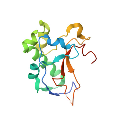
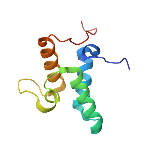
Ultrahigh specificity in a network of computationally designed protein-interaction pairs.

Netzer, R.Listov, D.Lipsh, R.Dym, O.Albeck, S.Knop, O.Kleanthous, C.Fleishman, S.J.

(2018) Nat Commun 9: 5286-5286

  • DOI: https://doi.org/10.1038/s41467-018-07722-9
  • Primary Citation of Related Structures:  
    6ER6, 6ERE

  • PubMed Abstract: 

    Protein networks in all organisms comprise homologous interacting pairs. In these networks, some proteins are specific, interacting with one or a few binding partners, whereas others are multispecific and bind a range of targets. We describe an algorithm that starts from an interacting pair and designs dozens of new pairs with diverse backbone conformations at the binding site as well as new binding orientations and sequences. Applied to a high-affinity bacterial pair, the algorithm results in 18 new ones, with cognate affinities from pico- to micromolar. Three pairs exhibit 3-5 orders of magnitude switch in specificity relative to the wild type, whereas others are multispecific, collectively forming a protein-interaction network. Crystallographic analysis confirms design accuracy, including in new backbones and polar interactions. Preorganized polar interaction networks are responsible for high specificity, thus defining design principles that can be applied to program synthetic cellular interaction networks of desired affinity and specificity.


  • Organizational Affiliation

    Department of Biomolecular Sciences, Weizmann Institute of Science, 7610001, Rehovot, Israel.


Macromolecules
Find similar proteins by:  (by identity cutoff)  |  3D Structure
Entity ID: 1
MoleculeChains Sequence LengthOrganismDetailsImage
Endonuclease colEdes7A [auth B]134Escherichia coliMutation(s): 0 
Entity Groups  
Sequence Clusters30% Identity50% Identity70% Identity90% Identity95% Identity100% Identity
Sequence Annotations
Expand
  • Reference Sequence
Find similar proteins by:  (by identity cutoff)  |  3D Structure
Entity ID: 2
MoleculeChains Sequence LengthOrganismDetailsImage
immunity Imdes7B [auth A]93Escherichia coliMutation(s): 0 
Entity Groups  
Sequence Clusters30% Identity50% Identity70% Identity90% Identity95% Identity100% Identity
Sequence Annotations
Expand
  • Reference Sequence
Experimental Data & Validation

Experimental Data

  • Method: X-RAY DIFFRACTION
  • Resolution: 1.60 Å
  • R-Value Free: 0.241 
  • R-Value Work: 0.197 
  • R-Value Observed: 0.199 
  • Space Group: P 21 21 21
Unit Cell:
Length ( Å )Angle ( ˚ )
a = 39.34α = 90
b = 56.297β = 90
c = 101.977γ = 90
Software Package:
Software NamePurpose
REFMACrefinement
HKL-2000data reduction
SCALAdata scaling
PHASERphasing

Structure Validation

View Full Validation Report


Entry History 

Deposition Data

Revision History  (Full details and data files)

  • Version 1.0: 2019-01-30
    Type: Initial release
  • Version 1.1: 2019-04-10
    Changes: Data collection, Derived calculations
  • Version 1.2: 2020-02-12
    Changes: Database references, Structure summary
  • Version 1.3: 2020-02-19
    Changes: Database references
  • Version 2.0: 2020-02-26
    Type: Coordinate replacement
    Reason: Occupancy of atoms on special symmetry positions
    Changes: Advisory, Atomic model, Author supporting evidence, Data collection, Derived calculations, Refinement description, Structure summary
  • Version 2.1: 2024-01-17
    Changes: Data collection, Database references, Refinement description