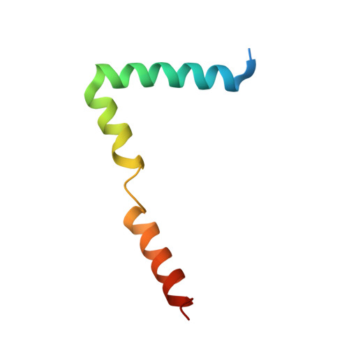
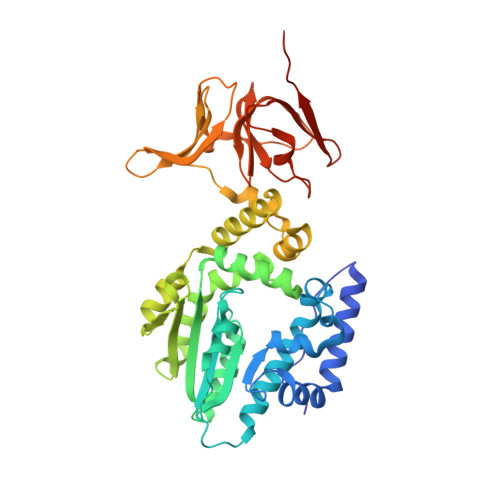
6EPH

Structure of the epsilon_1 / zeta_1 antitoxin toxin system from Neisseria gonorrhoeae in complex with UNAM.


Experimental Data Snapshot

  • Method: X-RAY DIFFRACTION
  • Resolution: 2.70 Å
  • R-Value Free: 0.245 
  • R-Value Work: 0.220 
  • R-Value Observed: 0.221 

wwPDB Validation   3D Report Full Report


Ligand Structure Quality Assessment 


This is version 1.2 of the entry. See complete history


Literature

The ng_ zeta 1 toxin of the gonococcal epsilon/zeta toxin/antitoxin system drains precursors for cell wall synthesis.

Rocker, A.Peschke, M.Kittila, T.Sakson, R.Brieke, C.Meinhart, A.

(2018) Nat Commun 9: 1686-1686

  • DOI: https://doi.org/10.1038/s41467-018-03652-8
  • Primary Citation of Related Structures:  
    6EPG, 6EPH, 6EPI

  • PubMed Abstract: 

    Bacterial toxin-antitoxin complexes are emerging as key players modulating bacterial physiology as activation of toxins induces stasis or programmed cell death by interference with vital cellular processes. Zeta toxins, which are prevalent in many bacterial genomes, were shown to interfere with cell wall formation by perturbing peptidoglycan synthesis in Gram-positive bacteria. Here, we characterize the epsilon/zeta toxin-antitoxin (TA) homologue from the Gram-negative pathogen Neisseria gonorrhoeae termed ng_ɛ1 / ng_ζ1. Contrary to previously studied streptococcal epsilon/zeta TA systems, ng_ɛ1 has an epsilon-unrelated fold and ng_ζ1 displays broader substrate specificity and phosphorylates multiple UDP-activated sugars that are precursors of peptidoglycan and lipopolysaccharide synthesis. Moreover, the phosphorylation site is different from the streptococcal zeta toxins, resulting in a different interference with cell wall synthesis. This difference most likely reflects adaptation to the individual cell wall composition of Gram-negative and Gram-positive organisms but also the distinct involvement of cell wall components in virulence.


  • Organizational Affiliation

    Department of Biomolecular Mechanisms, Max Planck Institute for Medical Research, Jahnstr. 29, 69120, Heidelberg, Germany.


Macromolecules
Find similar proteins by:  (by identity cutoff)  |  3D Structure
Entity ID: 1
MoleculeChains Sequence LengthOrganismDetailsImage
Epsilon_1 antitoxin
A, C, E, G
61Neisseria gonorrhoeaeMutation(s): 0 
UniProt
Find proteins for D5K9E3 (Neisseria gonorrhoeae)
Explore D5K9E3 
Go to UniProtKB:  D5K9E3
Entity Groups  
Sequence Clusters30% Identity50% Identity70% Identity90% Identity95% Identity100% Identity
UniProt GroupD5K9E3
Sequence Annotations
Expand
  • Reference Sequence
Find similar proteins by:  (by identity cutoff)  |  3D Structure
Entity ID: 2
MoleculeChains Sequence LengthOrganismDetailsImage
Zeta_1 toxin
B, D, F, H
401Neisseria gonorrhoeaeMutation(s): 0 
UniProt
Find proteins for D5K9G7 (Neisseria gonorrhoeae)
Explore D5K9G7 
Go to UniProtKB:  D5K9G7
Entity Groups  
Sequence Clusters30% Identity50% Identity70% Identity90% Identity95% Identity100% Identity
UniProt GroupD5K9G7
Sequence Annotations
Expand
  • Reference Sequence
Small Molecules
Ligands 3 Unique
IDChains Name / Formula / InChI Key2D Diagram3D Interactions
EPZ
Query on EPZ

Download Ideal Coordinates CCD File 
I [auth B],
N [auth D],
R [auth F],
V [auth H]
(2R)-2-{[(2R,3R,4R,5S,6R)-3-(acetylamino)-2-{[(S)-{[(R)-{[(2R,3S,4R,5R)-5-(2,4-dioxo-3,4-dihydropyrimidin-1(2H)-yl)-3,4-dihydroxytetrahydrofuran-2-yl]methoxy}(hydroxy)phosphoryl]oxy}(hydroxy)phosphoryl]oxy}-5-hydroxy-6-(hydroxymethyl)tetrahydro-2H-pyran-4-yl]oxy}propanoic acid
C20 H31 N3 O19 P2
NQBRVZNDBBMBLJ-MQTLHLSBSA-N
FLC
Query on FLC

Download Ideal Coordinates CCD File 
Q [auth D],
U [auth F]
CITRATE ANION
C6 H5 O7
KRKNYBCHXYNGOX-UHFFFAOYSA-K
SO4
Query on SO4

Download Ideal Coordinates CCD File 
J [auth B]
K [auth B]
L [auth B]
M [auth B]
O [auth D]
J [auth B],
K [auth B],
L [auth B],
M [auth B],
O [auth D],
P [auth D],
S [auth F],
T [auth F],
W [auth H],
X [auth H],
Y [auth H],
Z [auth H]
SULFATE ION
O4 S
QAOWNCQODCNURD-UHFFFAOYSA-L
Experimental Data & Validation

Experimental Data

  • Method: X-RAY DIFFRACTION
  • Resolution: 2.70 Å
  • R-Value Free: 0.245 
  • R-Value Work: 0.220 
  • R-Value Observed: 0.221 
  • Space Group: P 1 21 1
Unit Cell:
Length ( Å )Angle ( ˚ )
a = 80.29α = 90
b = 148.18β = 93.88
c = 124.4γ = 90
Software Package:
Software NamePurpose
REFMACrefinement
XDSdata reduction
XSCALEdata scaling
Cootmodel building
REFMACphasing

Structure Validation

View Full Validation Report


Ligand Structure Quality Assessment 


Entry History 

Deposition Data

Revision History  (Full details and data files)

  • Version 1.0: 2018-05-02
    Type: Initial release
  • Version 1.1: 2018-05-09
    Changes: Data collection, Database references
  • Version 1.2: 2024-01-17
    Changes: Data collection, Database references, Refinement description