6EEL

Crystal Structure of Myoferlin C2A with divalent cation


Experimental Data Snapshot

  • Method: X-RAY DIFFRACTION
  • Resolution: 1.93 Å
  • R-Value Free: 0.232 
  • R-Value Work: 0.181 
  • R-Value Observed: 0.186 

wwPDB Validation   3D Report Full Report


This is version 1.3 of the entry. See complete history


Literature

Structural Basis for the Distinct Membrane Binding Activity of the Homologous C2A Domains of Myoferlin and Dysferlin.

Harsini, F.M.Bui, A.A.Rice, A.M.Chebrolu, S.Fuson, K.L.Turtoi, A.Bradberry, M.Chapman, E.R.Sutton, R.B.

(2019) J Mol Biol 431: 2112-2126

  • DOI: https://doi.org/10.1016/j.jmb.2019.04.006
  • Primary Citation of Related Structures:  
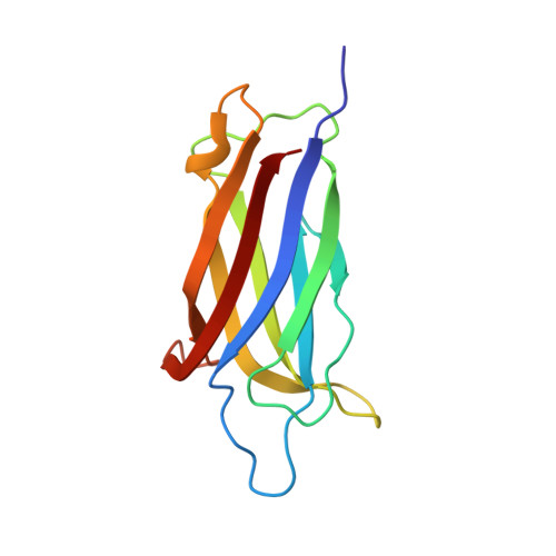
    6EEL

  • PubMed Abstract: 

    Dysferlin has been implicated in acute membrane repair processes, whereas myoferlin's activity is maximal during the myoblast fusion stage of early skeletal muscle cell development. Both proteins are similar in size and domain structure; however, despite the overall similarity, myoferlin's known physiological functions do not overlap with those of dysferlin. Here we present for the first time the X-ray crystal structure of human myoferlin C2A to 1.9 Å resolution bound to two divalent cations, and compare its three-dimensional structure and membrane binding activities to that of dysferlin C2A. We find that while dysferlin C2A binds membranes in a Ca 2+ -dependent manner, Ca 2+ binding was the rate-limiting kinetic step for this interaction. Myoferlin C2A, on the other hand, binds two calcium ions with an affinity 3-fold lower than that of dysferlin C2A; and, surprisingly, myoferlin C2A binds only marginally to phospholipid mixtures with a high fraction of phosphatidylserine.


  • Organizational Affiliation

    Department of Cell Physiology and Molecular Biophysics, Texas Tech University Health Sciences Center, Lubbock, TX, 79430, USA; Center for Membrane Protein Research, Texas Tech University Health Sciences Center, Lubbock, TX, 79430, USA.


Macromolecules
Find similar proteins by:  (by identity cutoff)  |  3D Structure
Entity ID: 1
MoleculeChains Sequence LengthOrganismDetailsImage
Myoferlin
A, B, C
129Homo sapiensMutation(s): 0 
Gene Names: MYOFFER1L3KIAA1207
Membrane Entity: Yes 
UniProt & NIH Common Fund Data Resources
Find proteins for Q9NZM1 (Homo sapiens)
Explore Q9NZM1 
Go to UniProtKB:  Q9NZM1
PHAROS:  Q9NZM1
GTEx:  ENSG00000138119 
Entity Groups  
Sequence Clusters30% Identity50% Identity70% Identity90% Identity95% Identity100% Identity
UniProt GroupQ9NZM1
Sequence Annotations
Expand
  • Reference Sequence
Experimental Data & Validation

Experimental Data

  • Method: X-RAY DIFFRACTION
  • Resolution: 1.93 Å
  • R-Value Free: 0.232 
  • R-Value Work: 0.181 
  • R-Value Observed: 0.186 
  • Space Group: P 21 21 21
Unit Cell:
Length ( Å )Angle ( ˚ )
a = 47.35α = 90
b = 83.11β = 90
c = 94.28γ = 90
Software Package:
Software NamePurpose
DENZOdata reduction
SCALAdata scaling
REFMACrefinement
PDB_EXTRACTdata extraction
PHASERphasing

Structure Validation

View Full Validation Report


Entry History & Funding Information

Deposition Data


Funding OrganizationLocationGrant Number
National Institutes of Health/National Institute of Arthritis and Musculoskeletal and Skin Diseases (NIH/NIAMS)United StatesAR063634

Revision History  (Full details and data files)

  • Version 1.0: 2019-05-22
    Type: Initial release
  • Version 1.1: 2019-06-05
    Changes: Data collection, Database references
  • Version 1.2: 2019-12-11
    Changes: Author supporting evidence
  • Version 1.3: 2023-10-11
    Changes: Data collection, Database references, Derived calculations, Refinement description