


Crystal structure of Leishmania major dihydroorotate dehydrogenase mutant H174A in complex with orotate
Reis, R.A.G., Pinheiro, M.P., de Souza, A.L., Hunter, W.N., Nonato, M.C.To be published.
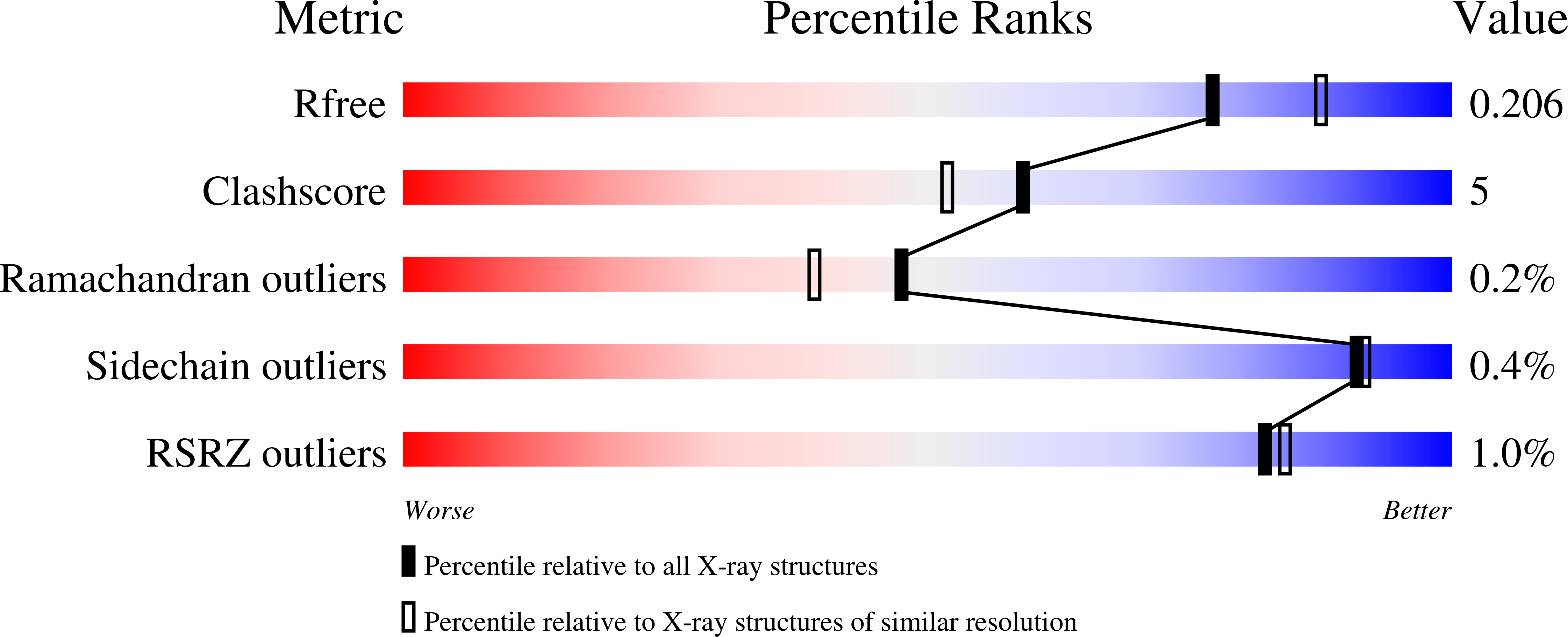
Experimental Data Snapshot
Entity ID: 1 | |||||
|---|---|---|---|---|---|
| Molecule | Chains | Sequence Length | Organism | Details | Image |
| Dihydroorotate dehydrogenase (fumarate) | 354 | Leishmania major | Mutation(s): 1 Gene Names: DHODH, LMJF_16_0530 EC: 1.3.98.1 | 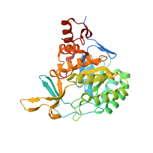 | |
UniProt | |||||
Find proteins for Q4QEW7 (Leishmania major) Explore Q4QEW7 Go to UniProtKB: Q4QEW7 | |||||
Entity Groups | |||||
| Sequence Clusters | 30% Identity50% Identity70% Identity90% Identity95% Identity100% Identity | ||||
| UniProt Group | Q4QEW7 | ||||
Sequence AnnotationsExpand | |||||
| |||||
| Ligands 5 Unique | |||||
|---|---|---|---|---|---|
| ID | Chains | Name / Formula / InChI Key | 2D Diagram | 3D Interactions | |
| FMN Query on FMN | C [auth A], K [auth B] | FLAVIN MONONUCLEOTIDE C17 H21 N4 O9 P FVTCRASFADXXNN-SCRDCRAPSA-N |  | ||
| ORO Query on ORO | D [auth A], L [auth B] | OROTIC ACID C5 H4 N2 O4 PXQPEWDEAKTCGB-UHFFFAOYSA-N |  | ||
| SO4 Query on SO4 | H [auth A] I [auth A] J [auth A] N [auth B] O [auth B] | SULFATE ION O4 S QAOWNCQODCNURD-UHFFFAOYSA-L |  | ||
| GOL Query on GOL | E [auth A], F [auth A], G [auth A], M [auth B] | GLYCEROL C3 H8 O3 PEDCQBHIVMGVHV-UHFFFAOYSA-N |  | ||
| NI Query on NI | Q [auth B] | NICKEL (II) ION Ni VEQPNABPJHWNSG-UHFFFAOYSA-N |  | ||
| Length ( Å ) | Angle ( ˚ ) |
|---|---|
| a = 144.279 | α = 90 |
| b = 144.279 | β = 90 |
| c = 69.913 | γ = 120 |
| Software Name | Purpose |
|---|---|
| Aimless | data scaling |
| MOLREP | phasing |
| REFMAC | refinement |
| PDB_EXTRACT | data extraction |
| Funding Organization | Location | Grant Number |
|---|---|---|
| Sao Paulo Research Foundation (FAPESP) | Brazil | 11/23504-9 |
| Sao Paulo Research Foundation (FAPESP) | Brazil | 07/08703-0 |
RCSB PDB (citation) is hosted by
RCSB PDB is a member of the

