6DV2

Crystal Structure of Human Mitochondrial Trifunctional Protein


Experimental Data Snapshot

  • Method: X-RAY DIFFRACTION
  • Resolution: 3.60 Å
  • R-Value Free: 0.289 
  • R-Value Work: 0.244 
  • R-Value Observed: 0.244 

wwPDB Validation   3D Report Full Report


This is version 1.5 of the entry. See complete history


Literature

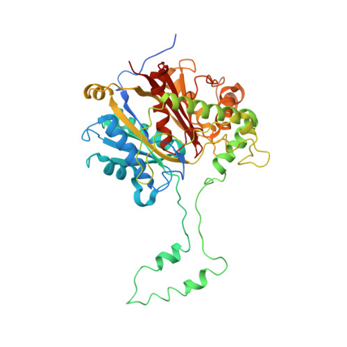
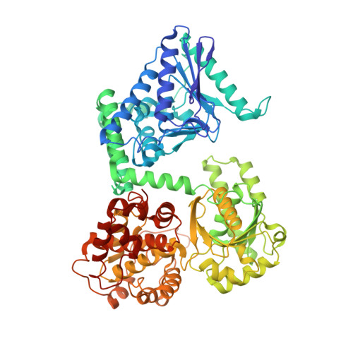
Crystal structure of human mitochondrial trifunctional protein, a fatty acid beta-oxidation metabolon.

Xia, C.Fu, Z.Battaile, K.P.Kim, J.P.

(2019) Proc Natl Acad Sci U S A 116: 6069-6074

  • DOI: https://doi.org/10.1073/pnas.1816317116
  • Primary Citation of Related Structures:  
    6DV2

  • PubMed Abstract: 

    Membrane-bound mitochondrial trifunctional protein (TFP) catalyzes β-oxidation of long chain fatty acyl-CoAs, employing 2-enoyl-CoA hydratase (ECH), 3-hydroxyl-CoA dehydrogenase (HAD), and 3-ketothiolase (KT) activities consecutively. Inherited deficiency of TFP is a recessive genetic disease, manifesting in hypoketotic hypoglycemia, cardiomyopathy, and sudden death. We have determined the crystal structure of human TFP at 3.6-Å resolution. The biological unit of the protein is α 2 β 2 The overall structure of the heterotetramer is the same as that observed by cryo-EM methods. The two β-subunits make a tightly bound homodimer at the center, and two α-subunits are bound to each side of the β 2 dimer, creating an arc, which binds on its concave side to the mitochondrial innermembrane. The catalytic residues in all three active sites are arranged similarly to those of the corresponding, soluble monofunctional enzymes. A structure-based, substrate channeling pathway from the ECH active site to the HAD and KT sites is proposed. The passage from the ECH site to the HAD site is similar to those found in the two bacterial TFPs. However, the passage from the HAD site to the KT site is unique in that the acyl-CoA intermediate can be transferred between the two sites by passing along the mitochondrial inner membrane using the hydrophobic nature of the acyl chain. The 3'-AMP-PPi moiety is guided by the positively charged residues located along the "ceiling" of the channel, suggesting that membrane integrity is an essential part of the channel and is required for the activity of the enzyme.


  • Organizational Affiliation

    Department of Biochemistry, Medical College of Wisconsin, Milwaukee, WI 53226.


Macromolecules
Find similar proteins by:  (by identity cutoff)  |  3D Structure
Entity ID: 1
MoleculeChains Sequence LengthOrganismDetailsImage
Trifunctional enzyme subunit beta, mitochondrial
A, B, C, D, E
A, B, C, D, E, F
457Homo sapiensMutation(s): 0 
Gene Names: HADHBMSTP029
EC: 2.3.1.16
Membrane Entity: Yes 
UniProt & NIH Common Fund Data Resources
Find proteins for P55084 (Homo sapiens)
Explore P55084 
Go to UniProtKB:  P55084
PHAROS:  P55084
GTEx:  ENSG00000138029 
Entity Groups  
Sequence Clusters30% Identity50% Identity70% Identity90% Identity95% Identity100% Identity
UniProt GroupP55084
Sequence Annotations
Expand
  • Reference Sequence
Find similar proteins by:  (by identity cutoff)  |  3D Structure
Entity ID: 2
MoleculeChains Sequence LengthOrganismDetailsImage
Trifunctional enzyme subunit alpha, mitochondrial
G, H, I, J, K
G, H, I, J, K, L
727Homo sapiensMutation(s): 0 
Gene Names: HADHAHADH
EC: 4.2.1.17 (PDB Primary Data), 1.1.1.211 (PDB Primary Data)
Membrane Entity: Yes 
UniProt & NIH Common Fund Data Resources
Find proteins for P40939 (Homo sapiens)
Explore P40939 
Go to UniProtKB:  P40939
PHAROS:  P40939
GTEx:  ENSG00000084754 
Entity Groups  
Sequence Clusters30% Identity50% Identity70% Identity90% Identity95% Identity100% Identity
UniProt GroupP40939
Sequence Annotations
Expand
  • Reference Sequence
Experimental Data & Validation

Experimental Data

  • Method: X-RAY DIFFRACTION
  • Resolution: 3.60 Å
  • R-Value Free: 0.289 
  • R-Value Work: 0.244 
  • R-Value Observed: 0.244 
  • Space Group: P 1 21 1
Unit Cell:
Length ( Å )Angle ( ˚ )
a = 136.536α = 90
b = 237.938β = 105.61
c = 141.319γ = 90
Software Package:
Software NamePurpose
CNSrefinement
SCALAdata scaling
PDB_EXTRACTdata extraction
MOSFLMdata reduction
PHASERphasing

Structure Validation

View Full Validation Report


Entry History & Funding Information

Deposition Data


Funding OrganizationLocationGrant Number
Department of Health & Human Services (HHS)United StatesGM29076

Revision History  (Full details and data files)

  • Version 1.0: 2018-09-26
    Type: Initial release
  • Version 1.1: 2019-03-06
    Changes: Data collection, Database references
  • Version 1.2: 2019-03-20
    Changes: Data collection, Database references
  • Version 1.3: 2019-04-10
    Changes: Data collection, Database references
  • Version 1.4: 2020-01-01
    Changes: Author supporting evidence
  • Version 1.5: 2023-10-11
    Changes: Data collection, Database references, Refinement description