6DQ0

sfGFP D133 mutated to 4-nitro-L-phenylalanine


Experimental Data Snapshot

  • Method: X-RAY DIFFRACTION
  • Resolution: 2.05 Å
  • R-Value Free: 0.219 
  • R-Value Work: 0.185 
  • R-Value Observed: 0.187 

Starting Model: experimental
View more details

wwPDB Validation   3D Report Full Report


This is version 1.3 of the entry. See complete history


Literature

Crystal structures of green fluorescent protein with the unnatural amino acid 4-nitro-L-phenylalanine.

Maurici, N.Savidge, N.Lee, B.U.Brewer, S.H.Phillips-Piro, C.M.

(2018) Acta Crystallogr F Struct Biol Commun 74: 650-655

  • DOI: https://doi.org/10.1107/S2053230X1801169X
  • Primary Citation of Related Structures:  
    6DQ0, 6DQ1

  • PubMed Abstract: 

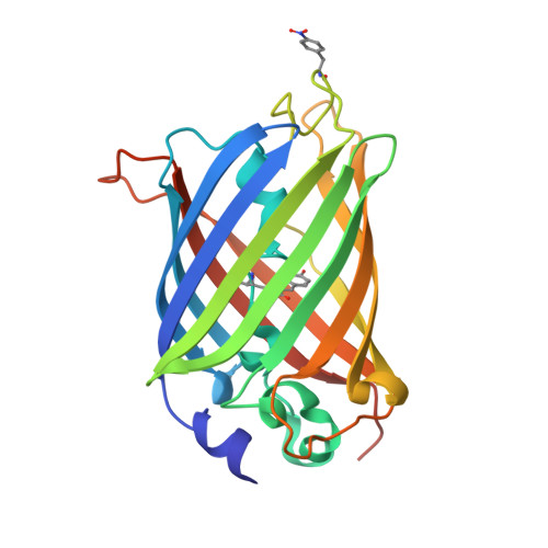
    The X-ray crystal structures of two superfolder green fluorescent protein (sfGFP) constructs containing a genetically incorporated spectroscopic reporter unnatural amino acid, 4-nitro-L-phenylalanine (pNO 2 F), at two unique sites in the protein have been determined. Amber codon-suppression methodology was used to site-specifically incorporate pNO 2 F at a solvent-accessible (Asp133) and a partially buried (Asn149) site in sfGFP. The Asp133pNO 2 F sfGFP construct crystallized with two molecules per asymmetric unit in space group P3 2 21 and the crystal structure was refined to 2.05 Å resolution. Crystals of Asn149pNO 2 F sfGFP contained one molecule of sfGFP per asymmetric unit in space group P4 1 22 and the structure was refined to 1.60 Å resolution. The alignment of Asp133pNO 2 F or Asn149pNO 2 F sfGFP with wild-type sfGFP resulted in small root-mean-square deviations, illustrating that these residues do not significantly alter the protein structure and supporting the use of pNO 2 F as an effective spectroscopic reporter of local protein structure and dynamics.


  • Organizational Affiliation

    Department of Chemistry, Franklin and Marshall College, PO Box 3003, Lancaster, PA 17604, USA.


Macromolecules
Find similar proteins by:  (by identity cutoff)  |  3D Structure
Entity ID: 1
MoleculeChains Sequence LengthOrganismDetailsImage
superfolder green fluorescent protein
A, B
237Aequorea victoriaMutation(s): 0 
UniProt
Find proteins for P42212 (Aequorea victoria)
Explore P42212 
Go to UniProtKB:  P42212
Entity Groups  
Sequence Clusters30% Identity50% Identity70% Identity90% Identity95% Identity100% Identity
UniProt GroupP42212
Sequence Annotations
Expand
  • Reference Sequence
Experimental Data & Validation

Experimental Data

  • Method: X-RAY DIFFRACTION
  • Resolution: 2.05 Å
  • R-Value Free: 0.219 
  • R-Value Work: 0.185 
  • R-Value Observed: 0.187 
  • Space Group: P 32 2 1
Unit Cell:
Length ( Å )Angle ( ˚ )
a = 97.72α = 90
b = 97.72β = 90
c = 154.872γ = 120
Software Package:
Software NamePurpose
PHENIXrefinement
HKL-2000data reduction
HKL-2000data scaling
PHASERphasing

Structure Validation

View Full Validation Report


Entry History & Funding Information

Deposition Data


Funding OrganizationLocationGrant Number
National Science Foundation (NSF, United States)United StatesCHE-1053946

Revision History  (Full details and data files)

  • Version 1.0: 2018-10-17
    Type: Initial release
  • Version 1.1: 2019-11-27
    Changes: Author supporting evidence
  • Version 1.2: 2023-10-11
    Changes: Data collection, Database references, Refinement description
  • Version 1.3: 2023-11-15
    Changes: Data collection