6DKU

Crystal structure of Myotis VP35 mutant of interferon inhibitory domain


Experimental Data Snapshot

  • Method: X-RAY DIFFRACTION
  • Resolution: 2.60 Å
  • R-Value Free: 0.223 
  • R-Value Work: 0.171 
  • R-Value Observed: 0.173 

wwPDB Validation   3D Report Full Report


This is version 1.3 of the entry. See complete history


Literature

Conservation of Structure and Immune Antagonist Functions of Filoviral VP35 Homologs Present in Microbat Genomes.

Edwards, M.R.Liu, H.Shabman, R.S.Ginell, G.M.Luthra, P.Ramanan, P.Keefe, L.J.Kollner, B.Amarasinghe, G.K.Taylor, D.J.Leung, D.W.Basler, C.F.

(2018) Cell Rep 24: 861-872.e6

  • DOI: https://doi.org/10.1016/j.celrep.2018.06.045
  • Primary Citation of Related Structures:  
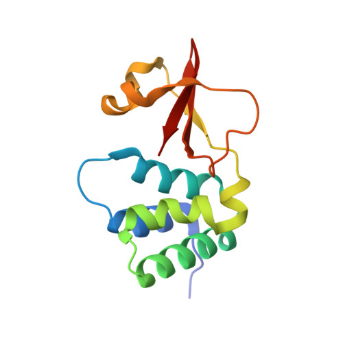
    6DKU

  • PubMed Abstract: 

    Non-retroviral integrated RNA viral sequences (NIRVs) potentially encoding ∼280 amino acid homologs to filovirus VP35 proteins are present across the Myotis genus of bats. These are estimated to have been maintained for ∼18 million years, indicating their co-option. To address the reasons for co-option, 16 Myotis VP35s were characterized in comparison to VP35s from the extant filoviruses Ebola virus and Marburg virus, in which VP35s play critical roles in immune evasion and RNA synthesis. The Myotis VP35s demonstrated a conserved suppression of innate immune signaling, albeit with reduced potency, in either human or Myotis cells. Their attenuation reflects a lack of dsRNA binding that in the filoviral VP35s correlates with potent suppression of interferon responses. Despite divergent function, evolution has preserved in Myotis the structure of the filoviral VP35s, indicating that this structure is critical for co-opted function, possibly as a regulator of innate immune signaling.


  • Organizational Affiliation

    Center for Microbial Pathogenesis, Institute for Biomedical Sciences, Georgia State University, Atlanta, GA 30303, USA.


Macromolecules
Find similar proteins by:  (by identity cutoff)  |  3D Structure
Entity ID: 1
MoleculeChains Sequence LengthOrganismDetailsImage
VP35128Myotis lucifugusMutation(s): 9 
UniProt
Find proteins for G9JKD1 (Myotis lucifugus)
Explore G9JKD1 
Go to UniProtKB:  G9JKD1
Entity Groups  
Sequence Clusters30% Identity50% Identity70% Identity90% Identity95% Identity100% Identity
UniProt GroupG9JKD1
Sequence Annotations
Expand
  • Reference Sequence
Experimental Data & Validation

Experimental Data

  • Method: X-RAY DIFFRACTION
  • Resolution: 2.60 Å
  • R-Value Free: 0.223 
  • R-Value Work: 0.171 
  • R-Value Observed: 0.173 
  • Space Group: P 32 2 1
Unit Cell:
Length ( Å )Angle ( ˚ )
a = 78.126α = 90
b = 78.126β = 90
c = 43.471γ = 120
Software Package:
Software NamePurpose
MOLREPphasing
PHENIXrefinement
PDB_EXTRACTdata extraction
autoPROCdata scaling
XDSdata reduction
XDSdata scaling

Structure Validation

View Full Validation Report


Entry History & Funding Information

Deposition Data


Funding OrganizationLocationGrant Number
National Institutes of Health/National Institute Of Allergy and Infectious Diseases (NIH/NIAID)United StatesAI109945

Revision History  (Full details and data files)

  • Version 1.0: 2018-07-18
    Type: Initial release
  • Version 1.1: 2018-08-08
    Changes: Data collection, Database references
  • Version 1.2: 2019-12-18
    Changes: Author supporting evidence
  • Version 1.3: 2023-10-11
    Changes: Data collection, Database references, Refinement description