6D9Q

The sulfate-bound crystal structure of HPRT (hypoxanthine phosphoribosyltransferase)


Experimental Data Snapshot

  • Method: X-RAY DIFFRACTION
  • Resolution: 2.06 Å
  • R-Value Free: 0.217 
  • R-Value Work: 0.183 
  • R-Value Observed: 0.184 

wwPDB Validation   3D Report Full Report


This is version 1.3 of the entry. See complete history


Literature

Evolution of (p)ppGpp-HPRT regulation through diversification of an allosteric oligomeric interaction.

Anderson, B.W.Liu, K.Wolak, C.Dubiel, K.She, F.Satyshur, K.A.Keck, J.L.Wang, J.D.

(2019) Elife 8

  • DOI: https://doi.org/10.7554/eLife.47534
  • Primary Citation of Related Structures:  
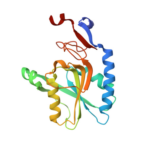
    6D9Q, 6D9R, 6D9S

  • PubMed Abstract: 

    The alarmone (p)ppGpp regulates diverse targets, yet its target specificity and evolution remain poorly understood. Here, we elucidate the mechanism by which basal (p)ppGpp inhibits the purine salvage enzyme HPRT by sharing a conserved motif with its substrate PRPP. Intriguingly, HPRT regulation by (p)ppGpp varies across organisms and correlates with HPRT oligomeric forms. (p)ppGpp-sensitive HPRT exists as a PRPP-bound dimer or an apo- and (p)ppGpp-bound tetramer, where a dimer-dimer interface triggers allosteric structural rearrangements to enhance (p)ppGpp inhibition. Loss of this oligomeric interface results in weakened (p)ppGpp regulation. Our results reveal an evolutionary principle whereby protein oligomerization allows evolutionary change to accumulate away from a conserved binding pocket to allosterically alter specificity of ligand interaction. This principle also explains how another (p)ppGpp target GMK is variably regulated across species. Since most ligands bind near protein interfaces, we propose that this principle extends to many other protein-ligand interactions.


  • Organizational Affiliation

    Department of Bacteriology, University of Wisconsin, Madison, United States.


Macromolecules
Find similar proteins by:  (by identity cutoff)  |  3D Structure
Entity ID: 1
MoleculeChains Sequence LengthOrganismDetailsImage
Hypoxanthine phosphoribosyltransferase
A, B, C, D
183Bacillus anthracisMutation(s): 0 
Gene Names: 
EC: 2.4.2.8
UniProt
Find proteins for B9ZW32 (Bacillus anthracis)
Explore B9ZW32 
Go to UniProtKB:  B9ZW32
Entity Groups  
Sequence Clusters30% Identity50% Identity70% Identity90% Identity95% Identity100% Identity
UniProt GroupB9ZW32
Sequence Annotations
Expand
  • Reference Sequence
Experimental Data & Validation

Experimental Data

  • Method: X-RAY DIFFRACTION
  • Resolution: 2.06 Å
  • R-Value Free: 0.217 
  • R-Value Work: 0.183 
  • R-Value Observed: 0.184 
  • Space Group: P 31 2 1
Unit Cell:
Length ( Å )Angle ( ˚ )
a = 82.597α = 90
b = 82.597β = 90
c = 242.416γ = 120
Software Package:
Software NamePurpose
PHENIXrefinement
HKL-2000data reduction
HKL-2000data scaling
PHASERphasing
ADSCdata collection

Structure Validation

View Full Validation Report


Entry History & Funding Information

Deposition Data


Funding OrganizationLocationGrant Number
National Institutes of Health/National Institute of General Medical Sciences (NIH/NIGMS)United StatesGM084003
National Institutes of Health/National Institute of General Medical Sciences (NIH/NIGMS)United StatesGM07215
National Science Foundation (NSF, United States)United StatesDGE-1256259

Revision History  (Full details and data files)

  • Version 1.0: 2019-05-01
    Type: Initial release
  • Version 1.1: 2019-11-13
    Changes: Database references
  • Version 1.2: 2019-11-27
    Changes: Author supporting evidence
  • Version 1.3: 2023-10-04
    Changes: Data collection, Database references, Refinement description