6D0Y

X-ray Crystal Structure of PGC-1beta C-terminus bound to the CBP80-CBP20 Cap Binding Complex


Experimental Data Snapshot

  • Method: X-RAY DIFFRACTION
  • Resolution: 2.68 Å
  • R-Value Free: 0.240 
  • R-Value Work: 0.181 
  • R-Value Observed: 0.185 

wwPDB Validation   3D Report Full Report


Ligand Structure Quality Assessment 


This is version 1.2 of the entry. See complete history


Literature

Transcriptional coactivator PGC-1 alpha contains a novel CBP80-binding motif that orchestrates efficient target gene expression.

Cho, H.Rambout, X.Gleghorn, M.L.Nguyen, P.Q.T.Phipps, C.R.Miyoshi, K.Myers, J.R.Kataoka, N.Fasan, R.Maquat, L.E.

(2018) Genes Dev 32: 555-567

  • DOI: https://doi.org/10.1101/gad.309773.117
  • Primary Citation of Related Structures:  
    6D0Y

  • PubMed Abstract: 

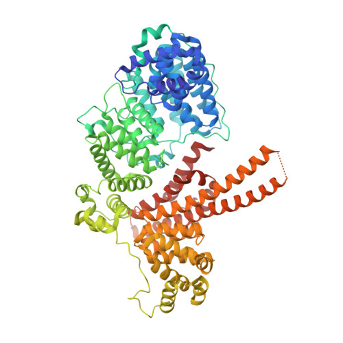
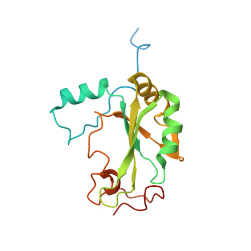
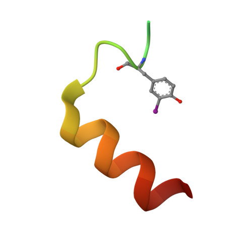
    Although peroxisome proliferator-activated receptor-γ (PPARγ) coactivator 1α (PGC-1α) is a well-established transcriptional coactivator for the metabolic adaptation of mammalian cells to diverse physiological stresses, the molecular mechanism by which it functions is incompletely understood. Here we used in vitro binding assays, X-ray crystallography, and immunoprecipitations of mouse myoblast cell lysates to define a previously unknown cap-binding protein 80 (CBP80)-binding motif (CBM) in the C terminus of PGC-1α. We show that the CBM, which consists of a nine-amino-acid α helix, is critical for the association of PGC-1α with CBP80 at the 5' cap of target transcripts. Results from RNA sequencing demonstrate that the PGC-1α CBM promotes RNA synthesis from promyogenic genes. Our findings reveal a new conduit between DNA-associated and RNA-associated proteins that functions in a cap-binding protein surveillance mechanism, without which efficient differentiation of myoblasts to myotubes fails to occur.


  • Organizational Affiliation

    Department of Biochemistry and Biophysics, School of Medicine and Dentistry, University of Rochester, Rochester, New York 14642, USA.


Macromolecules
Find similar proteins by:  (by identity cutoff)  |  3D Structure
Entity ID: 1
MoleculeChains Sequence LengthOrganismDetailsImage
Nuclear cap-binding protein subunit 1A [auth C]782Homo sapiensMutation(s): 0 
Gene Names: NCBP1CBP80NCBP
UniProt & NIH Common Fund Data Resources
Find proteins for Q09161 (Homo sapiens)
Explore Q09161 
Go to UniProtKB:  Q09161
PHAROS:  Q09161
GTEx:  ENSG00000136937 
Entity Groups  
Sequence Clusters30% Identity50% Identity70% Identity90% Identity95% Identity100% Identity
UniProt GroupQ09161
Sequence Annotations
Expand
  • Reference Sequence
Find similar proteins by:  (by identity cutoff)  |  3D Structure
Entity ID: 2
MoleculeChains Sequence LengthOrganismDetailsImage
Nuclear cap-binding protein subunit 2B [auth A]176Homo sapiensMutation(s): 0 
Gene Names: NCBP2CBP20PIG55
UniProt & NIH Common Fund Data Resources
Find proteins for P52298 (Homo sapiens)
Explore P52298 
Go to UniProtKB:  P52298
PHAROS:  P52298
GTEx:  ENSG00000114503 
Entity Groups  
Sequence Clusters30% Identity50% Identity70% Identity90% Identity95% Identity100% Identity
UniProt GroupP52298
Sequence Annotations
Expand
  • Reference Sequence
Find similar proteins by:  (by identity cutoff)  |  3D Structure
Entity ID: 3
MoleculeChains Sequence LengthOrganismDetailsImage
Peroxisome proliferator-activated receptor gamma coactivator 1-betaC [auth B]31Homo sapiensMutation(s): 0 
UniProt & NIH Common Fund Data Resources
Find proteins for Q86YN6 (Homo sapiens)
Explore Q86YN6 
Go to UniProtKB:  Q86YN6
PHAROS:  Q86YN6
GTEx:  ENSG00000155846 
Entity Groups  
Sequence Clusters30% Identity50% Identity70% Identity90% Identity95% Identity100% Identity
UniProt GroupQ86YN6
Sequence Annotations
Expand
  • Reference Sequence
Small Molecules
Ligands 2 Unique
IDChains Name / Formula / InChI Key2D Diagram3D Interactions
GTA
Query on GTA

Download Ideal Coordinates CCD File 
E [auth A]P1-7-METHYLGUANOSINE-P3-ADENOSINE-5',5'-TRIPHOSPHATE
C21 H30 N10 O17 P3
QQOHNVHGNZYSBP-XPWFQUROSA-O
MG
Query on MG

Download Ideal Coordinates CCD File 
D [auth C]MAGNESIUM ION
Mg
JLVVSXFLKOJNIY-UHFFFAOYSA-N
Modified Residues  1 Unique
IDChains TypeFormula2D DiagramParent
IYR
Query on IYR
C [auth B]L-PEPTIDE LINKINGC9 H10 I N O3TYR
Experimental Data & Validation

Experimental Data

  • Method: X-RAY DIFFRACTION
  • Resolution: 2.68 Å
  • R-Value Free: 0.240 
  • R-Value Work: 0.181 
  • R-Value Observed: 0.185 
  • Space Group: P 21 21 21
Unit Cell:
Length ( Å )Angle ( ˚ )
a = 78.106α = 90
b = 111.897β = 90
c = 124.795γ = 90
Software Package:
Software NamePurpose
Aimlessdata scaling
PHENIXrefinement
PDB_EXTRACTdata extraction
XDSdata reduction
PHENIXphasing

Structure Validation

View Full Validation Report


Ligand Structure Quality Assessment 


Entry History & Funding Information

Deposition Data


Funding OrganizationLocationGrant Number
National Institutes of Health/National Institute of General Medical Sciences (NIH/NIGMS)United StatesR01 GM59514

Revision History  (Full details and data files)

  • Version 1.0: 2018-04-25
    Type: Initial release
  • Version 1.1: 2018-05-09
    Changes: Data collection, Database references
  • Version 1.2: 2020-01-01
    Changes: Author supporting evidence