6CVL

Crystal structure of the Escherichia coli ATPgS-bound MetNI methionine ABC transporter in complex with its MetQ binding protein


Experimental Data Snapshot

  • Method: X-RAY DIFFRACTION
  • Resolution: 2.95 Å
  • R-Value Free: 0.226 
  • R-Value Work: 0.208 
  • R-Value Observed: 0.211 

wwPDB Validation   3D Report Full Report


Ligand Structure Quality Assessment 


This is version 1.2 of the entry. See complete history


Literature

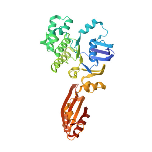
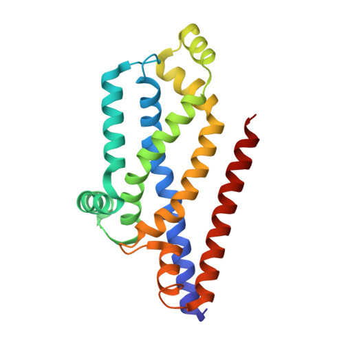
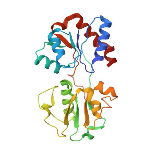
Noncanonical role for the binding protein in substrate uptake by the MetNI methionine ATP Binding Cassette (ABC) transporter.

Nguyen, P.T.Lai, J.Y.Lee, A.T.Kaiser, J.T.Rees, D.C.

(2018) Proc Natl Acad Sci U S A 115: E10596-E10604

  • DOI: https://doi.org/10.1073/pnas.1811003115
  • Primary Citation of Related Structures:  
    6CVL

  • PubMed Abstract: 

    The Escherichia coli methionine ABC transporter MetNI exhibits both high-affinity transport toward l-methionine and broad specificity toward methionine derivatives, including d-methionine. In this work, we characterize the transport of d-methionine derivatives by the MetNI transporter. Unexpectedly, the N229A substrate-binding deficient variant of the cognate binding protein MetQ was found to support high MetNI transport activity toward d-selenomethionine. We determined the crystal structure at 2.95 Å resolution of the ATPγS-bound MetNIQ complex in the outward-facing conformation with the N229A apo MetQ variant. This structure revealed conformational changes in MetQ providing substrate access through the binding protein to the transmembrane translocation pathway. MetQ likely mediates uptake of methionine derivatives through two mechanisms: in the methionine-bound form delivering substrate from the periplasm to the transporter (the canonical mechanism) and in the apo form by facilitating ligand binding when complexed to the transporter (the noncanonical mechanism). This dual role for substrate-binding proteins is proposed to provide a kinetic strategy for ABC transporters to transport both high- and low-affinity substrates present in a physiological concentration range.


  • Organizational Affiliation

    Division of Chemistry and Chemical Engineering, California Institute of Technology, Pasadena, CA 91125.


Macromolecules
Find similar proteins by:  (by identity cutoff)  |  3D Structure
Entity ID: 1
MoleculeChains Sequence LengthOrganismDetailsImage
MetN nucleotide-binding subunitA [auth C],
D
344Escherichia coli K-12Mutation(s): 2 
Gene Names: metNabcb0199JW0195
EC: 3.6.3
Membrane Entity: Yes 
UniProt
Find proteins for P30750 (Escherichia coli (strain K12))
Explore P30750 
Go to UniProtKB:  P30750
Entity Groups  
Sequence Clusters30% Identity50% Identity70% Identity90% Identity95% Identity100% Identity
UniProt GroupP30750
Sequence Annotations
Expand
  • Reference Sequence
Find similar proteins by:  (by identity cutoff)  |  3D Structure
Entity ID: 2
MoleculeChains Sequence LengthOrganismDetailsImage
MetI transmembrane subunitB [auth A],
C [auth B]
215Escherichia coli K-12Mutation(s): 0 
Gene Names: metIyaeEb0198JW0194
Membrane Entity: Yes 
UniProt
Find proteins for P31547 (Escherichia coli (strain K12))
Explore P31547 
Go to UniProtKB:  P31547
Entity Groups  
Sequence Clusters30% Identity50% Identity70% Identity90% Identity95% Identity100% Identity
UniProt GroupP31547
Sequence Annotations
Expand
  • Reference Sequence
Find similar proteins by:  (by identity cutoff)  |  3D Structure
Entity ID: 3
MoleculeChains Sequence LengthOrganismDetailsImage
MetQ periplasmic binding protein226Escherichia coli K-12Mutation(s): 1 
Gene Names: metQyaeCb0197JW0193
Membrane Entity: Yes 
UniProt
Find proteins for P28635 (Escherichia coli (strain K12))
Explore P28635 
Go to UniProtKB:  P28635
Entity Groups  
Sequence Clusters30% Identity50% Identity70% Identity90% Identity95% Identity100% Identity
UniProt GroupP28635
Sequence Annotations
Expand
  • Reference Sequence
Experimental Data & Validation

Experimental Data

  • Method: X-RAY DIFFRACTION
  • Resolution: 2.95 Å
  • R-Value Free: 0.226 
  • R-Value Work: 0.208 
  • R-Value Observed: 0.211 
  • Space Group: P 32 2 1
Unit Cell:
Length ( Å )Angle ( ˚ )
a = 107.96α = 90
b = 107.96β = 90
c = 354.52γ = 120
Software Package:
Software NamePurpose
PHENIXrefinement
XDSdata reduction
Aimlessdata scaling
PHASERphasing
DMphasing
PDB_EXTRACTdata extraction

Structure Validation

View Full Validation Report


Ligand Structure Quality Assessment 


Entry History & Funding Information

Deposition Data


Funding OrganizationLocationGrant Number
Howard Hughes Medical Institute (HHMI)United States--
Vietnam International Education DevelopmentViet Nam--

Revision History  (Full details and data files)

  • Version 1.0: 2018-11-14
    Type: Initial release
  • Version 1.1: 2019-11-20
    Changes: Advisory, Author supporting evidence
  • Version 1.2: 2022-04-27
    Changes: Advisory, Database references, Derived calculations