6BHY

Mouse Immunoglobulin G 2c Fc fragment with single GlcNAc

  • Classification: IMMUNE SYSTEM
  • Organism(s): Mus musculus
  • Expression System: Homo sapiens
  • Mutation(s): No 

  • Deposited: 2017-10-31 Released: 2018-02-14 
  • Deposition Author(s): Falconer, D., Barb, A.
  • Funding Organization(s): National Institutes of Health/National Institute of General Medical Sciences (NIH/NIGMS)

Experimental Data Snapshot

  • Method: X-RAY DIFFRACTION
  • Resolution: 2.04 Å
  • R-Value Free: 0.254 
  • R-Value Work: 0.210 
  • R-Value Observed: 0.213 

wwPDB Validation   3D Report Full Report


Ligand Structure Quality Assessment 


This is version 1.3 of the entry. See complete history


Literature

Mouse IgG2c Fc loop residues promote greater receptor-binding affinity than mouse IgG2b or human IgG1.

Falconer, D.J.Barb, A.W.

(2018) PLoS One 13: e0192123-e0192123

  • DOI: https://doi.org/10.1371/journal.pone.0192123
  • Primary Citation of Related Structures:  
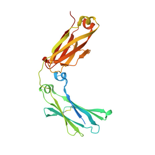
    6BHQ, 6BHY

  • PubMed Abstract: 

    The structures of non-human antibodies are largely unstudied despite the potential for the identification of alternative structural motifs and physical properties that will benefit a basic understanding of protein and immune system evolution as well as highlight unexplored motifs to improve therapeutic monoclonal antibody. Here we probe the structure and receptor-binding properties of the mouse IgG2c crystallizable fragment (Fc) to compare to mouse IgG2b and human IgG1 Fcs. Models of mIgG2c Fc determined by x-ray crystallography with a complex-type biantennary (to 2.05 Å) or a truncated (1)GlcNAc asparagine-linked (N)-glycan attached (to 2.04 Å) show differences in key regions related to mouse Fc γ receptor IV (mFcγRIV) binding. Mouse IgG2c forms different non-bonded interactions between the BC, DE and FG loops than the highly-conserved mIgG2b and binds to FcγRIV with 4.7-fold greater affinity in the complex-type glycoform. Secondary structural elements surrounding the Asn297 site of glycosylation form longer beta strands in the truncated mIgG2c Fc glycoform when compared to mIgG2c with the complex-type N-glycan. Solution NMR spectroscopy of the N-linked (1)GlcNAc residues show differences between mIgG2b, 2c and hIgG1 Fc that correlate to receptor binding affinity. Mutations targeting differences in mIgG2 DE and FG loops decreased affinity of mIgG2c for FcγRIV and increased affinity of mIgG2b. Changes in NMR spectra of the mutant Fc proteins mirrored these changes in affinity. Our studies identified structural and functional differences in highly conserved molecules that were not predicted from primary sequence comparison.


  • Organizational Affiliation

    Roy J. Carver Department of Biochemistry, Biophysics and Molecular Biology Iowa State University, Ames, IA, United States of America.


Macromolecules
Find similar proteins by:  (by identity cutoff)  |  3D Structure
Entity ID: 1
MoleculeChains Sequence LengthOrganismDetailsImage
Igh protein
A, B
238Mus musculusMutation(s): 0 
Gene Names: Igh
UniProt
Find proteins for Q58E56 (Mus musculus)
Explore Q58E56 
Go to UniProtKB:  Q58E56
Entity Groups  
Sequence Clusters30% Identity50% Identity70% Identity90% Identity95% Identity100% Identity
UniProt GroupQ58E56
Sequence Annotations
Expand
  • Reference Sequence
Experimental Data & Validation

Experimental Data

  • Method: X-RAY DIFFRACTION
  • Resolution: 2.04 Å
  • R-Value Free: 0.254 
  • R-Value Work: 0.210 
  • R-Value Observed: 0.213 
  • Space Group: C 1 2 1
Unit Cell:
Length ( Å )Angle ( ˚ )
a = 103.233α = 90
b = 89.215β = 132.2
c = 69.851γ = 90
Software Package:
Software NamePurpose
PHENIXrefinement
JBluIce-EPICSdata collection
HKL-2000data reduction
HKL-2000data scaling
PHENIXphasing

Structure Validation

View Full Validation Report


Ligand Structure Quality Assessment 


Entry History & Funding Information

Deposition Data


Funding OrganizationLocationGrant Number
National Institutes of Health/National Institute of General Medical Sciences (NIH/NIGMS)United StatesGM115489

Revision History  (Full details and data files)

  • Version 1.0: 2018-02-14
    Type: Initial release
  • Version 1.1: 2020-01-01
    Changes: Author supporting evidence
  • Version 1.2: 2020-07-29
    Type: Remediation
    Reason: Carbohydrate remediation
    Changes: Data collection, Derived calculations, Structure summary
  • Version 1.3: 2023-10-04
    Changes: Data collection, Database references, Refinement description, Structure summary