6BDZ

ADAM10 Extracellular Domain Bound by the 11G2 Fab


Experimental Data Snapshot

  • Method: X-RAY DIFFRACTION
  • Resolution: 3.10 Å
  • R-Value Free: 0.317 
  • R-Value Work: 0.261 
  • R-Value Observed: 0.267 

wwPDB Validation   3D Report Full Report


This is version 2.0 of the entry. See complete history


Literature

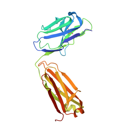
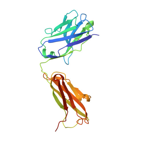
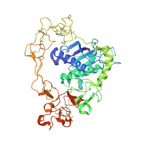
Structural Basis for Regulated Proteolysis by the alpha-Secretase ADAM10.

Seegar, T.C.M.Killingsworth, L.B.Saha, N.Meyer, P.A.Patra, D.Zimmerman, B.Janes, P.W.Rubinstein, E.Nikolov, D.B.Skiniotis, G.Kruse, A.C.Blacklow, S.C.

(2017) Cell 171: 1638-1648.e7

  • DOI: https://doi.org/10.1016/j.cell.2017.11.014
  • Primary Citation of Related Structures:  
    6BDZ, 6BE6

  • PubMed Abstract: 

    Cleavage of membrane-anchored proteins by ADAM (a disintegrin and metalloproteinase) endopeptidases plays a key role in a wide variety of biological signal transduction and protein turnover processes. Among ADAM family members, ADAM10 stands out as particularly important because it is both responsible for regulated proteolysis of Notch receptors and catalyzes the non-amyloidogenic α-secretase cleavage of the Alzheimer's precursor protein (APP). We present here the X-ray crystal structure of the ADAM10 ectodomain, which, together with biochemical and cellular studies, reveals how access to the enzyme active site is regulated. The enzyme adopts an unanticipated architecture in which the C-terminal cysteine-rich domain partially occludes the enzyme active site, preventing unfettered substrate access. Binding of a modulatory antibody to the cysteine-rich domain liberates the catalytic domain from autoinhibition, enhancing enzymatic activity toward a peptide substrate. Together, these studies reveal a mechanism for regulation of ADAM activity and offer a roadmap for its modulation.


  • Organizational Affiliation

    Department of Biological Chemistry and Molecular Pharmacology, Harvard Medical School, Boston, MA 02115, USA.


Macromolecules
Find similar proteins by:  (by identity cutoff)  |  3D Structure
Entity ID: 1
MoleculeChains Sequence LengthOrganismDetailsImage
11G2 Fab Light ChainA [auth L]215Mus musculusMutation(s): 0 
Entity Groups  
Sequence Clusters30% Identity50% Identity70% Identity90% Identity95% Identity100% Identity
Sequence Annotations
Expand
  • Reference Sequence
Find similar proteins by:  (by identity cutoff)  |  3D Structure
Entity ID: 2
MoleculeChains Sequence LengthOrganismDetailsImage
11G2 Fab Heavy ChainB [auth H]226Mus musculusMutation(s): 0 
Entity Groups  
Sequence Clusters30% Identity50% Identity70% Identity90% Identity95% Identity100% Identity
Sequence Annotations
Expand
  • Reference Sequence
Find similar proteins by:  (by identity cutoff)  |  3D Structure
Entity ID: 3
MoleculeChains Sequence LengthOrganismDetailsImage
Disintegrin and metalloproteinase domain-containing protein 10C [auth A]443Homo sapiensMutation(s): 0 
Gene Names: ADAM10KUZMADM
EC: 3.4.24.81
UniProt & NIH Common Fund Data Resources
Find proteins for O14672 (Homo sapiens)
Explore O14672 
Go to UniProtKB:  O14672
PHAROS:  O14672
GTEx:  ENSG00000137845 
Entity Groups  
Sequence Clusters30% Identity50% Identity70% Identity90% Identity95% Identity100% Identity
UniProt GroupO14672
Sequence Annotations
Expand
  • Reference Sequence
Oligosaccharides

Help

Entity ID: 4
MoleculeChains Length2D Diagram Glycosylation3D Interactions
alpha-D-mannopyranose-(1-6)-alpha-D-mannopyranose-(1-4)-2-acetamido-2-deoxy-beta-D-glucopyranose-(1-4)-2-acetamido-2-deoxy-beta-D-glucopyranoseD [auth B]4N-Glycosylation
Glycosylation Resources
GlyTouCan:  G04854NQ
GlyCosmos:  G04854NQ
GlyGen:  G04854NQ
Entity ID: 5
MoleculeChains Length2D Diagram Glycosylation3D Interactions
alpha-D-mannopyranose-(1-4)-2-acetamido-2-deoxy-beta-D-glucopyranose-(1-4)-2-acetamido-2-deoxy-beta-D-glucopyranoseE [auth C]3N-Glycosylation
Glycosylation Resources
GlyTouCan:  G62182OO
GlyCosmos:  G62182OO
GlyGen:  G62182OO
Experimental Data & Validation

Experimental Data

  • Method: X-RAY DIFFRACTION
  • Resolution: 3.10 Å
  • R-Value Free: 0.317 
  • R-Value Work: 0.261 
  • R-Value Observed: 0.267 
  • Space Group: C 2 2 21
Unit Cell:
Length ( Å )Angle ( ˚ )
a = 79.23α = 90
b = 97.58β = 90
c = 262.72γ = 90
Software Package:
Software NamePurpose
PHENIXrefinement
XDSdata scaling
PHENIXphasing

Structure Validation

View Full Validation Report


Entry History & Funding Information

Deposition Data


Funding OrganizationLocationGrant Number
National Institutes of Health/National Institute of General Medical Sciences (NIH/NIGMS)United StatesP41 GM103403

Revision History  (Full details and data files)

  • Version 1.0: 2017-12-27
    Type: Initial release
  • Version 1.1: 2020-01-01
    Changes: Author supporting evidence
  • Version 2.0: 2020-07-29
    Type: Remediation
    Reason: Carbohydrate remediation
    Changes: Advisory, Atomic model, Data collection, Derived calculations, Structure summary