6B0J

Crystal structure of Ps i-CgsB in complex with k-i-k-neocarrahexaose


Experimental Data Snapshot

  • Method: X-RAY DIFFRACTION
  • Resolution: 2.50 Å
  • R-Value Free: 0.234 
  • R-Value Work: 0.214 
  • R-Value Observed: 0.215 

wwPDB Validation   3D Report Full Report


Ligand Structure Quality Assessment 


This is version 2.1 of the entry. See complete history


Literature

The Molecular Basis of Polysaccharide Sulfatase Activity and a Nomenclature for Catalytic Subsites in this Class of Enzyme.

Hettle, A.G.Vickers, C.Robb, C.S.Liu, F.Withers, S.G.Hehemann, J.H.Boraston, A.B.

(2018) Structure 26: 747

  • DOI: https://doi.org/10.1016/j.str.2018.03.012
  • Primary Citation of Related Structures:  
    6B0J, 6B0K, 6B1V, 6BIA

  • PubMed Abstract: 

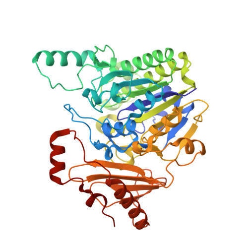
    Sulfatases play a biologically important role by cleaving sulfate groups from molecules. They can be identified on the basis of signature sequences within their primary structures, and the largest family, S1, has predictable features that contribute specifically to the recognition and catalytic removal of sulfate groups. However, despite advances in the prediction and understanding of S1 sulfatases, a major question regards the molecular determinants that drive substrate recognition beyond the targeted sulfate group. Here, through analysis of an endo-4S-ι-carrageenan sulfatase (PsS1_19A) from Pseudoalteromonas sp. PS47, particularly X-ray crystal structures in complex with intact substrates, we show that specific recognition of the substrate leaving group components, in this case carbohydrate, provides the enzyme with specificity for its substrate. On the basis of these results we propose a catalytic subsite nomenclature that we anticipate will form a general foundation for understanding and describing the molecular basis of substrate recognition by sulfatases.


  • Organizational Affiliation

    Department of Biochemistry and Microbiology, University of Victoria, Victoria, BC V8W 3P6, Canada.


Macromolecules
Find similar proteins by:  (by identity cutoff)  |  3D Structure
Entity ID: 1
MoleculeChains Sequence LengthOrganismDetailsImage
Iota-carrageenan sulfatase
A, B, C
452PseudoalteromonasMutation(s): 0 
Entity Groups  
Sequence Clusters30% Identity50% Identity70% Identity90% Identity95% Identity100% Identity
Sequence Annotations
Expand
  • Reference Sequence
Oligosaccharides

Help

Entity ID: 2
MoleculeChains Length2D Diagram Glycosylation3D Interactions
4-O-sulfo-beta-D-galactopyranose-(1-4)-3,6-anhydro-2-O-sulfo-alpha-D-galactopyranose-(1-3)-4-O-sulfo-beta-D-galactopyranose
D, F, G, H
3N/A
Glycosylation Resources
GlyTouCan:  G47184WN
GlyCosmos:  G47184WN
Entity ID: 3
MoleculeChains Length2D Diagram Glycosylation3D Interactions
3,6-anhydro-2-O-sulfo-alpha-D-galactopyranose-(1-3)-4-O-sulfo-beta-D-galactopyranose
E
2N/A
Glycosylation Resources
GlyTouCan:  G58187OK
GlyCosmos:  G58187OK
Small Molecules
Ligands 6 Unique
IDChains Name / Formula / InChI Key2D Diagram3D Interactions
G4S
Query on G4S

Download Ideal Coordinates CCD File 
J [auth A],
Q [auth B],
W [auth C]
4-O-sulfo-beta-D-galactopyranose
C6 H12 O9 S
LOTQRUGOUKUSEY-DGPNFKTASA-N
ARG
Query on ARG

Download Ideal Coordinates CCD File 
AA [auth C],
O [auth A]
ARGININE
C6 H15 N4 O2
ODKSFYDXXFIFQN-BYPYZUCNSA-O
9RN
Query on 9RN

Download Ideal Coordinates CCD File 
K [auth A]
L [auth A]
R [auth B]
S [auth B]
T [auth B]
K [auth A],
L [auth A],
R [auth B],
S [auth B],
T [auth B],
X [auth C],
Y [auth C]
3,6-anhydro-alpha-D-galactopyranose
C6 H10 O5
DCQFFOLNJVGHLW-RDQKPOQOSA-N
TRS
Query on TRS

Download Ideal Coordinates CCD File 
N [auth A],
U [auth B]
2-AMINO-2-HYDROXYMETHYL-PROPANE-1,3-DIOL
C4 H12 N O3
LENZDBCJOHFCAS-UHFFFAOYSA-O
EDO
Query on EDO

Download Ideal Coordinates CCD File 
M [auth A],
Z [auth C]
1,2-ETHANEDIOL
C2 H6 O2
LYCAIKOWRPUZTN-UHFFFAOYSA-N
CA
Query on CA

Download Ideal Coordinates CCD File 
I [auth A],
P [auth B],
V [auth C]
CALCIUM ION
Ca
BHPQYMZQTOCNFJ-UHFFFAOYSA-N
Experimental Data & Validation

Experimental Data

  • Method: X-RAY DIFFRACTION
  • Resolution: 2.50 Å
  • R-Value Free: 0.234 
  • R-Value Work: 0.214 
  • R-Value Observed: 0.215 
  • Space Group: P 43 21 2
Unit Cell:
Length ( Å )Angle ( ˚ )
a = 123.39α = 90
b = 123.39β = 90
c = 240.11γ = 90
Software Package:
Software NamePurpose
REFMACrefinement
MOSFLMdata reduction
SCALAdata scaling
PHASERphasing
PDB_EXTRACTdata extraction

Structure Validation

View Full Validation Report


Ligand Structure Quality Assessment 


Entry History 

Deposition Data

Revision History  (Full details and data files)

  • Version 1.0: 2018-03-14
    Type: Initial release
  • Version 1.1: 2018-05-09
    Changes: Data collection, Database references
  • Version 1.2: 2018-05-16
    Changes: Data collection, Database references
  • Version 2.0: 2020-07-29
    Type: Remediation
    Reason: Carbohydrate remediation
    Changes: Atomic model, Data collection, Derived calculations, Structure summary
  • Version 2.1: 2023-10-04
    Changes: Data collection, Database references, Derived calculations, Refinement description, Structure summary