6ANV

Crystal structure of anti-CRISPR protein AcrF1


Experimental Data Snapshot

  • Method: X-RAY DIFFRACTION
  • Resolution: 2.27 Å
  • R-Value Free: 0.223 
  • R-Value Work: 0.165 
  • R-Value Observed: 0.168 

wwPDB Validation   3D Report Full Report


This is version 2.1 of the entry. See complete history


Literature

Cryo-EM Structures Reveal Mechanism and Inhibition of DNA Targeting by a CRISPR-Cas Surveillance Complex.

Guo, T.W.Bartesaghi, A.Yang, H.Falconieri, V.Rao, P.Merk, A.Eng, E.T.Raczkowski, A.M.Fox, T.Earl, L.A.Patel, D.J.Subramaniam, S.

(2017) Cell 171: 414-426.e12

  • DOI: https://doi.org/10.1016/j.cell.2017.09.006
  • Primary Citation of Related Structures:  
    6ANV, 6ANW, 6B44, 6B45, 6B46, 6B47, 6B48

  • PubMed Abstract: 

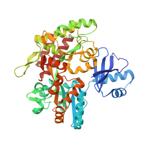
    Prokaryotic cells possess CRISPR-mediated adaptive immune systems that protect them from foreign genetic elements, such as invading viruses. A central element of this immune system is an RNA-guided surveillance complex capable of targeting non-self DNA or RNA for degradation in a sequence- and site-specific manner analogous to RNA interference. Although the complexes display considerable diversity in their composition and architecture, many basic mechanisms underlying target recognition and cleavage are highly conserved. Using cryoelectron microscopy (cryo-EM), we show that the binding of target double-stranded DNA (dsDNA) to a type I-F CRISPR system yersinia (Csy) surveillance complex leads to large quaternary and tertiary structural changes in the complex that are likely necessary in the pathway leading to target dsDNA degradation by a trans-acting helicase-nuclease. Comparison of the structure of the surveillance complex before and after dsDNA binding, or in complex with three virally encoded anti-CRISPR suppressors that inhibit dsDNA binding, reveals mechanistic details underlying target recognition and inhibition.


  • Organizational Affiliation

    Laboratory of Cell Biology, Center for Cancer Research, National Cancer Institute, NIH, Bethesda, MD 20892, USA.


Macromolecules
Find similar proteins by:  (by identity cutoff)  |  3D Structure
Entity ID: 1
MoleculeChains Sequence LengthOrganismDetailsImage
anti-CRISPR protein AcrF1 fused with C-terminal MBP tag
A, B
452Pseudomonas phage JBD30Escherichia coli O157:H7
This entity is chimeric
Mutation(s): 4 
Gene Names: JBD30_035malEZ5632ECs5017
UniProt
Find proteins for P0AEX9 (Escherichia coli (strain K12))
Explore P0AEX9 
Go to UniProtKB:  P0AEX9
Find proteins for L7P7M1 (Pseudomonas phage JBD30)
Explore L7P7M1 
Go to UniProtKB:  L7P7M1
Entity Groups  
Sequence Clusters30% Identity50% Identity70% Identity90% Identity95% Identity100% Identity
UniProt GroupsL7P7M1P0AEX9
Sequence Annotations
Expand
  • Reference Sequence
Oligosaccharides

Help

Entity ID: 2
MoleculeChains Length2D Diagram Glycosylation3D Interactions
alpha-D-glucopyranose-(1-4)-alpha-D-glucopyranose-(1-4)-alpha-D-glucopyranose-(1-4)-alpha-D-glucopyranose
C, D
4N/A
Glycosylation Resources
GlyTouCan:  G87171PZ
GlyCosmos:  G87171PZ
GlyGen:  G87171PZ
Small Molecules
Ligands 4 Unique
IDChains Name / Formula / InChI Key2D Diagram3D Interactions
MES
Query on MES

Download Ideal Coordinates CCD File 
E [auth A],
N [auth B]
2-(N-MORPHOLINO)-ETHANESULFONIC ACID
C6 H13 N O4 S
SXGZJKUKBWWHRA-UHFFFAOYSA-N
PGE
Query on PGE

Download Ideal Coordinates CCD File 
F [auth A],
O [auth B],
P [auth B]
TRIETHYLENE GLYCOL
C6 H14 O4
ZIBGPFATKBEMQZ-UHFFFAOYSA-N
PEG
Query on PEG

Download Ideal Coordinates CCD File 
J [auth A]
K [auth A]
L [auth A]
M [auth A]
U [auth B]
J [auth A],
K [auth A],
L [auth A],
M [auth A],
U [auth B],
V [auth B],
W [auth B]
DI(HYDROXYETHYL)ETHER
C4 H10 O3
MTHSVFCYNBDYFN-UHFFFAOYSA-N
EDO
Query on EDO

Download Ideal Coordinates CCD File 
G [auth A]
H [auth A]
I [auth A]
Q [auth B]
R [auth B]
G [auth A],
H [auth A],
I [auth A],
Q [auth B],
R [auth B],
S [auth B],
T [auth B]
1,2-ETHANEDIOL
C2 H6 O2
LYCAIKOWRPUZTN-UHFFFAOYSA-N
Biologically Interesting Molecules (External Reference) 1 Unique
Experimental Data & Validation

Experimental Data

  • Method: X-RAY DIFFRACTION
  • Resolution: 2.27 Å
  • R-Value Free: 0.223 
  • R-Value Work: 0.165 
  • R-Value Observed: 0.168 
  • Space Group: P 1 21 1
Unit Cell:
Length ( Å )Angle ( ˚ )
a = 87.389α = 90
b = 63.245β = 93.21
c = 96.733γ = 90
Software Package:
Software NamePurpose
PHENIXrefinement
HKL-2000data scaling
PDB_EXTRACTdata extraction
PHASERphasing

Structure Validation

View Full Validation Report


Entry History & Funding Information

Deposition Data


Funding OrganizationLocationGrant Number
National Institutes of Health/National Institute on Drug Abuse (NIH/NIDA)United StatesGM104962
Memorial Sloan-Kettering Cancer Center Core GrantUnited StatesP30CA008748

Revision History  (Full details and data files)

  • Version 1.0: 2017-10-25
    Type: Initial release
  • Version 1.1: 2019-12-11
    Changes: Author supporting evidence
  • Version 2.0: 2020-07-29
    Type: Remediation
    Reason: Carbohydrate remediation
    Changes: Atomic model, Data collection, Derived calculations, Non-polymer description, Structure summary
  • Version 2.1: 2023-10-04
    Changes: Data collection, Database references, Refinement description, Structure summary