6AID

Structural insights into the unique polylactate degrading mechanism of Thermobifida alba cutinase


Experimental Data Snapshot

  • Method: X-RAY DIFFRACTION
  • Resolution: 1.30 Å
  • R-Value Free: 0.165 
  • R-Value Work: 0.133 
  • R-Value Observed: 0.135 

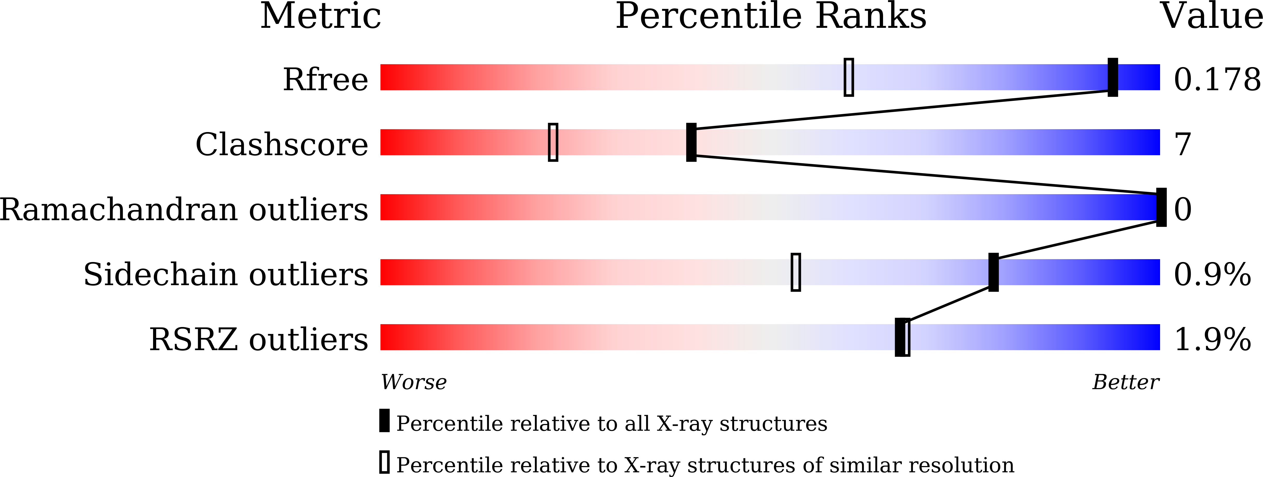
wwPDB Validation   3D Report Full Report


This is version 2.0 of the entry. See complete history


Literature

Structural insights into the unique polylactate-degrading mechanism of Thermobifida alba cutinase.

Kitadokoro, K.Kakara, M.Matsui, S.Osokoshi, R.Thumarat, U.Kawai, F.Kamitani, S.

(2019) FEBS J 286: 2087-2098

  • DOI: https://doi.org/10.1111/febs.14781
  • Primary Citation of Related Structures:  
    6AID

  • PubMed Abstract: 

    Cutinases are enzymes known to degrade polyester-type plastics. Est119, a plastic-degrading type of cutinase from Thermobifida alba AHK119 (herein called Ta_cut), shows a broad substrate specificity toward polyesters, and can degrade substrates including polylactic acid (PLA). However, the PLA-degrading mechanism of cutinases is still poorly understood. Here, we report the structure complexes of cutinase with ethyl lactate (EL), the constitutional unit. From this complex structure, the electron density maps clearly showed one lactate (LAC) and one EL occupying different positions in the active site cleft. The binding mode of EL is assumed to show a figure prior to reaction and LAC is an after-reaction product. These complex structures demonstrate the role of active site residues in the esterase reaction and substrate recognition. The complex structures were compared with other documented complex structures of cutinases and with the structure of PETase from Ideonella sakaiensis. The amino acid residues involved in substrate interaction are highly conserved among these enzymes. Thus, mapping the precise interactions in the Ta_cut and EL complex will pave the way for understanding the plastic-degrading mechanism of cutinases and suggest ways of creating more potent enzymes by structural protein engineering.


  • Organizational Affiliation

    Department of Biomolecular Engineering, Graduate School of Science and Technology, Kyoto Institute of Technology, Japan.


Macromolecules
Find similar proteins by:  (by identity cutoff)  |  3D Structure
Entity ID: 1
MoleculeChains Sequence LengthOrganismDetailsImage
Esterase278Thermobifida albaMutation(s): 0 
Gene Names: est2
UniProt
Find proteins for F7IX06 (Thermobifida alba)
Explore F7IX06 
Go to UniProtKB:  F7IX06
Entity Groups  
Sequence Clusters30% Identity50% Identity70% Identity90% Identity95% Identity100% Identity
UniProt GroupF7IX06
Sequence Annotations
Expand
  • Reference Sequence
Small Molecules
Ligands 4 Unique
IDChains Name / Formula / InChI Key2D Diagram3D Interactions
9YL
Query on 9YL

Download Ideal Coordinates CCD File 
E [auth A]ethyl (2R)-2-oxidanylpropanoate
C5 H10 O3
LZCLXQDLBQLTDK-SCSAIBSYSA-N
LAC
Query on LAC

Download Ideal Coordinates CCD File 
H [auth A]LACTIC ACID
C3 H6 O3
JVTAAEKCZFNVCJ-UWTATZPHSA-N
CA
Query on CA

Download Ideal Coordinates CCD File 
B [auth A],
C [auth A],
D [auth A]
CALCIUM ION
Ca
BHPQYMZQTOCNFJ-UHFFFAOYSA-N
NA
Query on NA

Download Ideal Coordinates CCD File 
F [auth A],
G [auth A]
SODIUM ION
Na
FKNQFGJONOIPTF-UHFFFAOYSA-N
Experimental Data & Validation

Experimental Data

  • Method: X-RAY DIFFRACTION
  • Resolution: 1.30 Å
  • R-Value Free: 0.165 
  • R-Value Work: 0.133 
  • R-Value Observed: 0.135 
  • Space Group: P 21 21 21
Unit Cell:
Length ( Å )Angle ( ˚ )
a = 42.265α = 90
b = 68.86β = 90
c = 78.594γ = 90
Software Package:
Software NamePurpose
REFMACrefinement
XDSdata reduction
XSCALEdata scaling
PHASERphasing

Structure Validation

View Full Validation Report


Entry History 

Deposition Data

Revision History  (Full details and data files)

  • Version 1.0: 2019-02-27
    Type: Initial release
  • Version 1.1: 2019-06-12
    Changes: Data collection, Database references
  • Version 2.0: 2023-11-15
    Changes: Atomic model, Data collection, Database references, Derived calculations, Refinement description