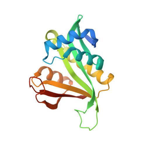
6AG4

Crystal structure of Ard1 N-terminal acetyltransferase H88A/E127A mutant from Sulfolobus solfataricus


Experimental Data Snapshot

  • Method: X-RAY DIFFRACTION
  • Resolution: 2.26 Å
  • R-Value Free: 0.229 
  • R-Value Work: 0.163 
  • R-Value Observed: 0.169 

wwPDB Validation   3D Report Full Report


Ligand Structure Quality Assessment 


This is version 1.2 of the entry. See complete history


Literature

Adaptation of thermophilic acetyltransferase to a water-mediated catalytic mechanism.

Chang, Y.Y.Hagawa, S.Hsu, C.H.

(2020) Chem Commun (Camb) 56: 10537-10540

  • DOI: https://doi.org/10.1039/d0cc04305b
  • Primary Citation of Related Structures:  
    6AG4, 6AG5

  • PubMed Abstract: 

    The common mechanism of N-acetyltransferases (NATs) is a water-mediated catalysis, which is not conducive to thermophilic acetyltransferases. The crystal structure of SsArd1 shows an ordered catalytic water molecule in a trap formed by the residues H88 and E127. Structure-guided mutagenesis, kinetic studies and MD simulation indicated that the turnover rates of H88A, E127A and H88A/E127A mutants were low, but that of the H88E/E127H mutant could be restored to the level of the wild type.


  • Organizational Affiliation

    Department of Agricultural Chemistry, National Taiwan University, Taipei 10617, Taiwan. andyhsu@ntu.edu.tw.


Macromolecules
Find similar proteins by:  (by identity cutoff)  |  3D Structure
Entity ID: 1
MoleculeChains Sequence LengthOrganismDetailsImage
N-alpha-acetyltransferase173Saccharolobus solfataricus P2Mutation(s): 2 
Gene Names: SsArd1
EC: 2.3.1.255 (PDB Primary Data), 2.3.1.258 (PDB Primary Data)
UniProt
Find proteins for Q980R9 (Saccharolobus solfataricus (strain ATCC 35092 / DSM 1617 / JCM 11322 / P2))
Explore Q980R9 
Go to UniProtKB:  Q980R9
Entity Groups  
Sequence Clusters30% Identity50% Identity70% Identity90% Identity95% Identity100% Identity
UniProt GroupQ980R9
Sequence Annotations
Expand
  • Reference Sequence
Experimental Data & Validation

Experimental Data

  • Method: X-RAY DIFFRACTION
  • Resolution: 2.26 Å
  • R-Value Free: 0.229 
  • R-Value Work: 0.163 
  • R-Value Observed: 0.169 
  • Space Group: P 21 21 21
Unit Cell:
Length ( Å )Angle ( ˚ )
a = 42.335α = 90
b = 53.148β = 90
c = 74.505γ = 90
Software Package:
Software NamePurpose
PHENIXrefinement
HKL-2000data reduction
HKL-2000data scaling
PHASERphasing

Structure Validation

View Full Validation Report


Ligand Structure Quality Assessment 


Entry History & Funding Information

Deposition Data


Funding OrganizationLocationGrant Number
Ministry of Science and Technology (Taiwan)Taiwan103-2113-M-002 -009 -MY2

Revision History  (Full details and data files)

  • Version 1.0: 2019-08-14
    Type: Initial release
  • Version 1.1: 2020-09-23
    Changes: Database references
  • Version 1.2: 2023-11-22
    Changes: Data collection, Database references, Refinement description