6Z65

Crystal structure of NAD kinase 1 from Listeria monocytogenes in complex with a di-adenosine derivative


Experimental Data Snapshot

  • Method: X-RAY DIFFRACTION
  • Resolution: 1.97 Å
  • R-Value Free: 0.228 
  • R-Value Work: 0.202 
  • R-Value Observed: 0.204 

wwPDB Validation   3D Report Full Report


Ligand Structure Quality Assessment 


This is version 1.1 of the entry. See complete history


Literature

New Chemical Probe Targeting Bacterial NAD Kinase.

Clement, D.A.Leseigneur, C.Gelin, M.Coelho, D.Huteau, V.Lionne, C.Labesse, G.Dussurget, O.Pochet, S.

(2020) Molecules 25

  • DOI: https://doi.org/10.3390/molecules25214893
  • Primary Citation of Related Structures:  
    6Z61, 6Z64, 6Z65

  • PubMed Abstract: 

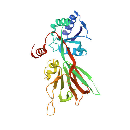
    Nicotinamide adenine dinucleotide (NAD) kinases are essential and ubiquitous enzymes involved in the tight regulation of NAD/nicotinamide adenine dinucleotide phosphate (NADP) levels in many metabolic pathways. Consequently, they represent promising therapeutic targets in cancer and antibacterial treatments. We previously reported diadenosine derivatives as NAD kinase inhibitors with bactericidal activities on Staphylococcus aureus . Among them, one compound (namely NKI1 ) was found effective in vivo in a mouse infection model. With the aim to gain detailed knowledge about the selectivity and mechanism of action of this lead compound, we planned to develop a chemical probe that could be used in affinity-based chemoproteomic approaches. Here, we describe the first functionalized chemical probe targeting a bacterial NAD kinase. Aminoalkyl functional groups were introduced on NKI1 for further covalent coupling to an activated Sepharose TM matrix. Inhibitory properties of functionalized NKI1 derivatives together with X-ray characterization of their complexes with the NAD kinase led to identify candidate compounds that are amenable to covalent coupling to a matrix.


  • Organizational Affiliation

    Institut Pasteur, Unité de Chimie et Biocatalyse, UMR3523 CNRS, 75015 Paris, France.


Macromolecules
Find similar proteins by:  (by identity cutoff)  |  3D Structure
Entity ID: 1
MoleculeChains Sequence LengthOrganismDetailsImage
NAD kinase 1272Listeria monocytogenes EGD-eMutation(s): 0 
Gene Names: nadK1lmo0968
EC: 2.7.1.23
UniProt
Find proteins for Q8Y8D7 (Listeria monocytogenes serovar 1/2a (strain ATCC BAA-679 / EGD-e))
Explore Q8Y8D7 
Go to UniProtKB:  Q8Y8D7
Entity Groups  
Sequence Clusters30% Identity50% Identity70% Identity90% Identity95% Identity100% Identity
UniProt GroupQ8Y8D7
Sequence Annotations
Expand
  • Reference Sequence
Small Molecules
Ligands 2 Unique
IDChains Name / Formula / InChI Key2D Diagram3D Interactions
Q9N (Subject of Investigation/LOI)
Query on Q9N

Download Ideal Coordinates CCD File 
C [auth A]~{N}-[[(2~{R},3~{S},4~{R},5~{R})-5-[8-[3-[[(2~{R},3~{S},4~{R},5~{R})-5-(6-aminopurin-9-yl)-3,4-bis(oxidanyl)oxolan-2-yl]methoxy]prop-1-ynyl]-6-azanyl-purin-9-yl]-3,4-bis(oxidanyl)oxolan-2-yl]methyl]-4-azanyl-butanamide
C27 H34 N12 O8
FSTDOBBDPHECFF-GMIOVBLFSA-N
CIT
Query on CIT

Download Ideal Coordinates CCD File 
B [auth A]CITRIC ACID
C6 H8 O7
KRKNYBCHXYNGOX-UHFFFAOYSA-N
Experimental Data & Validation

Experimental Data

  • Method: X-RAY DIFFRACTION
  • Resolution: 1.97 Å
  • R-Value Free: 0.228 
  • R-Value Work: 0.202 
  • R-Value Observed: 0.204 
  • Space Group: I 2 2 2
Unit Cell:
Length ( Å )Angle ( ˚ )
a = 63.019α = 90
b = 74.531β = 90
c = 118.739γ = 90
Software Package:
Software NamePurpose
PHENIXrefinement
XDSdata reduction
Aimlessdata scaling
PDB_EXTRACTdata extraction
PHENIXphasing

Structure Validation

View Full Validation Report


Ligand Structure Quality Assessment 


Entry History 

Deposition Data

Revision History  (Full details and data files)

  • Version 1.0: 2021-05-26
    Type: Initial release
  • Version 1.1: 2024-01-24
    Changes: Data collection, Database references, Refinement description