6YJN

Crystal structure of beta carbonic anhydrase from the pathogenic bacterium Burkholderia pseudomallei.


Experimental Data Snapshot

  • Method: X-RAY DIFFRACTION
  • Resolution: 2.70 Å
  • R-Value Free: 0.322 
  • R-Value Work: 0.213 
  • R-Value Observed: 0.219 

wwPDB Validation   3D Report Full Report


This is version 1.2 of the entry. See complete history


Literature

Crystal Structure of a Tetrameric Type II beta-Carbonic Anhydrase from the Pathogenic BacteriumBurkholderia pseudomallei.

Angeli, A.Ferraroni, M.Pinteala, M.Maier, S.S.Simionescu, B.C.Carta, F.Del Prete, S.Capasso, C.Supuran, C.T.

(2020) Molecules 25

  • DOI: https://doi.org/10.3390/molecules25102269
  • Primary Citation of Related Structures:  
    6YJN, 6YL7

  • PubMed Abstract: 

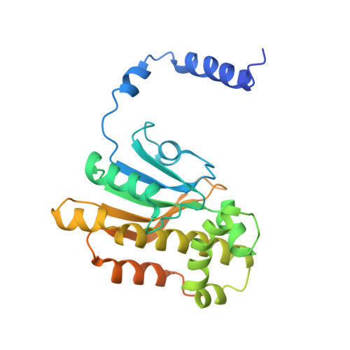
    Carbonic anhydrase (CA) is a zinc enzyme that catalyzes the reversible conversion of carbon dioxide to bicarbonate and proton. Currently, CA inhibitors are widely used as antiglaucoma, anticancer, and anti-obesity drugs and for the treatment of neurological disorders. Recently, the potential use of CA inhibitors to fight infections caused by protozoa, fungi, and bacteria has emerged as a new research line. In this article, the X-ray crystal structure of β-CA from Burkholderia pseudomallei was reported. The X-ray crystal structure of this new enzyme was solved at 2.7 Å resolution, revealing a tetrameric type II β-CA with a "closed" active site in which the zinc is tetrahedrally coordinated to Cys46, Asp48, His102, and Cys105. B. pseudomallei is known to encode at least two CAs, a β-CA, and a γ-CA. These proteins, playing a pivotal role in its life cycle and pathogenicity, offer a novel therapeutic opportunity to obtain antibiotics with a different mechanism of action. Furthermore, the new structure can provide a clear view of the β-CA mechanism of action and the possibility to find selective inhibitors for this class of CAs.


  • Organizational Affiliation

    NEUROFARBA Department, Sezione di Scienze Farmaceutiche, Via Ugo Schiff 6, Università degli Studi di Firenze, 50019 Sesto Fiorentino (Florence), Italy.


Macromolecules
Find similar proteins by:  (by identity cutoff)  |  3D Structure
Entity ID: 1
MoleculeChains Sequence LengthOrganismDetailsImage
Beta carbonic anhydrase256Burkholderia pseudomalleiMutation(s): 0 
Gene Names: can_1can_2BOC42_16450CXQ84_19645DF122_01465ERS013345_01543SAMEA1968934_03740
EC: 4.2.1.1
UniProt
Find proteins for Q63Y17 (Burkholderia pseudomallei (strain K96243))
Explore Q63Y17 
Go to UniProtKB:  Q63Y17
Entity Groups  
Sequence Clusters30% Identity50% Identity70% Identity90% Identity95% Identity100% Identity
UniProt GroupQ63Y17
Sequence Annotations
Expand
  • Reference Sequence
Small Molecules
Ligands 1 Unique
IDChains Name / Formula / InChI Key2D Diagram3D Interactions
ZN (Subject of Investigation/LOI)
Query on ZN

Download Ideal Coordinates CCD File 
B [auth A]ZINC ION
Zn
PTFCDOFLOPIGGS-UHFFFAOYSA-N
Experimental Data & Validation

Experimental Data

  • Method: X-RAY DIFFRACTION
  • Resolution: 2.70 Å
  • R-Value Free: 0.322 
  • R-Value Work: 0.213 
  • R-Value Observed: 0.219 
  • Space Group: P 64 2 2
Unit Cell:
Length ( Å )Angle ( ˚ )
a = 88.031α = 90
b = 88.031β = 90
c = 111.644γ = 120
Software Package:
Software NamePurpose
REFMACrefinement
Aimlessdata scaling
PDB_EXTRACTdata extraction
XDSdata reduction
MOLREPphasing

Structure Validation

View Full Validation Report


Entry History 

Deposition Data

Revision History  (Full details and data files)

  • Version 1.0: 2020-06-10
    Type: Initial release
  • Version 1.1: 2020-11-11
    Changes: Derived calculations
  • Version 1.2: 2024-01-24
    Changes: Data collection, Database references, Refinement description