6X1H

Crystal structure of a guanine nucleotide exchange factor (GEF) domain from the Orientia tsutsugamushi protein OtDUB


Experimental Data Snapshot

  • Method: X-RAY DIFFRACTION
  • Resolution: 2.91 Å
  • R-Value Free: 0.208 
  • R-Value Work: 0.166 
  • R-Value Observed: 0.168 

wwPDB Validation   3D Report Full Report


This is version 1.2 of the entry. See complete history


Literature

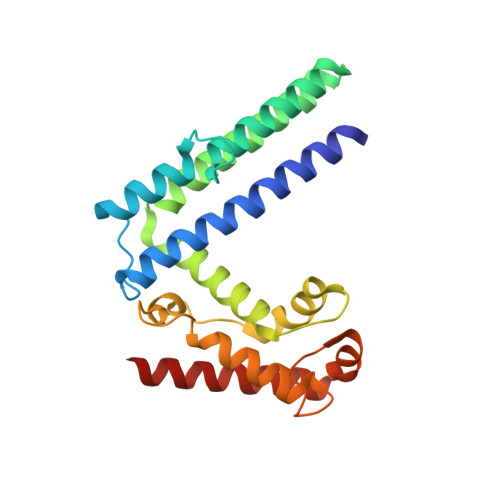
Crystal structure of a guanine nucleotide exchange factor encoded by the scrub typhus pathogen Orientia tsutsugamushi .

Lim, C.Berk, J.M.Blaise, A.Bircher, J.Koleske, A.J.Hochstrasser, M.Xiong, Y.

(2020) Proc Natl Acad Sci U S A 117: 30380-30390

  • DOI: https://doi.org/10.1073/pnas.2018163117
  • Primary Citation of Related Structures:  
    6X1G, 6X1H

  • PubMed Abstract: 

    Rho family GTPases regulate an array of cellular processes and are often modulated by pathogens to promote infection. Here, we identify a cryptic guanine nucleotide exchange factor (GEF) domain in the OtDUB protein encoded by the pathogenic bacterium Orientia tsutsugamushi A proteomics-based OtDUB interaction screen identified numerous potential host interactors, including the Rho GTPases Rac1 and Cdc42. We discovered a domain in OtDUB with Rac1/Cdc42 GEF activity (OtDUB GEF ), with higher activity toward Rac1 in vitro. While this GEF bears no obvious sequence similarity to known GEFs, crystal structures of OtDUB GEF alone (3.0 Å) and complexed with Rac1 (1.7 Å) reveal striking convergent evolution, with a unique topology, on a V-shaped bacterial GEF fold shared with other bacterial GEF domains. Structure-guided mutational analyses identified residues critical for activity and a mechanism for nucleotide displacement. Ectopic expression of OtDUB activates Rac1 preferentially in cells, and expression of the OtDUB GEF alone alters cell morphology. Cumulatively, this work reveals a bacterial GEF within the multifunctional OtDUB that co-opts host Rac1 signaling to induce changes in cytoskeletal structure.


  • Organizational Affiliation

    Department of Molecular Biophysics & Biochemistry, Yale University, New Haven, CT 06520.


Macromolecules
Find similar proteins by:  (by identity cutoff)  |  3D Structure
Entity ID: 1
MoleculeChains Sequence LengthOrganismDetailsImage
ULP_PROTEASE domain-containing protein219Orientia tsutsugamushi str. IkedaMutation(s): 0 
Gene Names: OTT_1962
UniProt
Find proteins for B3CVM3 (Orientia tsutsugamushi (strain Ikeda))
Explore B3CVM3 
Go to UniProtKB:  B3CVM3
Entity Groups  
Sequence Clusters30% Identity50% Identity70% Identity90% Identity95% Identity100% Identity
UniProt GroupB3CVM3
Sequence Annotations
Expand
  • Reference Sequence
Small Molecules
Ligands 1 Unique
IDChains Name / Formula / InChI Key2D Diagram3D Interactions
NI
Query on NI

Download Ideal Coordinates CCD File 
G [auth D]NICKEL (II) ION
Ni
VEQPNABPJHWNSG-UHFFFAOYSA-N
Experimental Data & Validation

Experimental Data

  • Method: X-RAY DIFFRACTION
  • Resolution: 2.91 Å
  • R-Value Free: 0.208 
  • R-Value Work: 0.166 
  • R-Value Observed: 0.168 
  • Space Group: P 63
Unit Cell:
Length ( Å )Angle ( ˚ )
a = 110.533α = 90
b = 110.533β = 90
c = 251.24γ = 120
Software Package:
Software NamePurpose
HKL-2000data scaling
REFMACrefinement
PDB_EXTRACTdata extraction
HKL-2000data reduction
PHASERphasing

Structure Validation

View Full Validation Report


Entry History & Funding Information

Deposition Data


Funding OrganizationLocationGrant Number
National Science Foundation (NSF, United States)United StatesDGE1122492

Revision History  (Full details and data files)

  • Version 1.0: 2020-11-25
    Type: Initial release
  • Version 1.1: 2020-12-16
    Changes: Database references
  • Version 1.2: 2023-10-18
    Changes: Data collection, Database references, Refinement description