6WUP

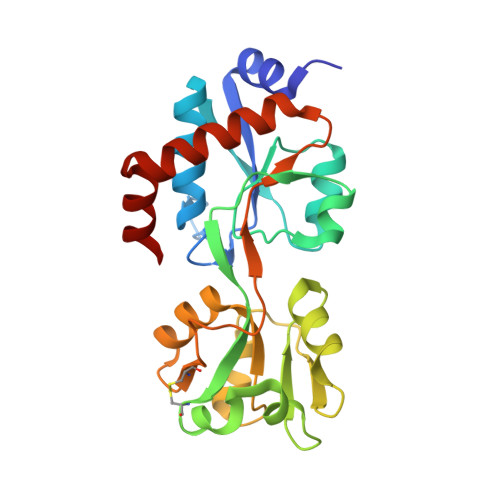
Crystal structure of an ancestral cyclohexadienyl dehydratase, AncCDT-5


Experimental Data Snapshot

  • Method: X-RAY DIFFRACTION
  • Resolution: 1.49 Å
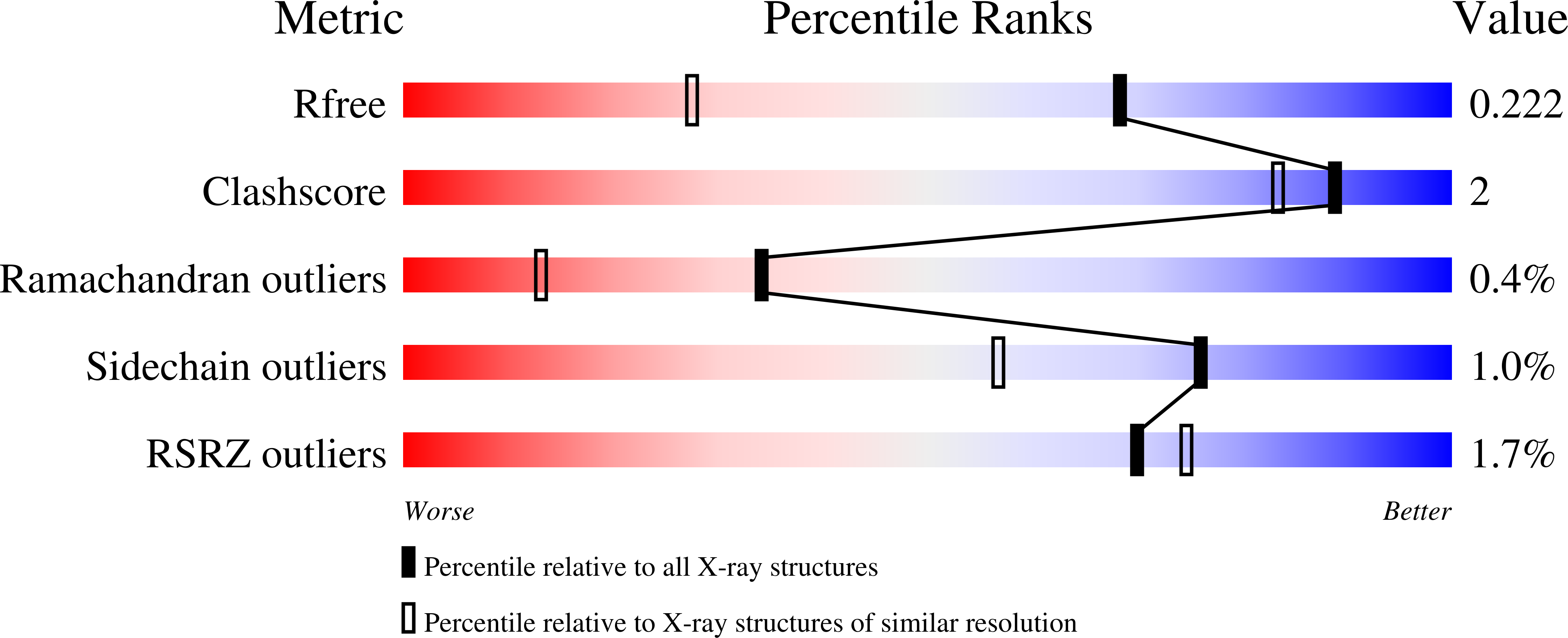
  • R-Value Free: 0.222 
  • R-Value Work: 0.198 
  • R-Value Observed: 0.199 

wwPDB Validation   3D Report Full Report


This is version 1.2 of the entry. See complete history


Literature

Altered conformational sampling along an evolutionary trajectory changes the catalytic activity of an enzyme.

Kaczmarski, J.A.Mahawaththa, M.C.Feintuch, A.Clifton, B.E.Adams, L.A.Goldfarb, D.Otting, G.Jackson, C.J.

(2020) Nat Commun 11: 5945-5945

  • DOI: https://doi.org/10.1038/s41467-020-19695-9
  • Primary Citation of Related Structures:  
    6WUP

  • PubMed Abstract: 

    Several enzymes are known to have evolved from non-catalytic proteins such as solute-binding proteins (SBPs). Although attention has been focused on how a binding site can evolve to become catalytic, an equally important question is: how do the structural dynamics of a binding protein change as it becomes an efficient enzyme? Here we performed a variety of experiments, including propargyl-DO3A-Gd(III) tagging and double electron-electron resonance (DEER) to study the rigid body protein dynamics of reconstructed evolutionary intermediates to determine how the conformational sampling of a protein changes along an evolutionary trajectory linking an arginine SBP to a cyclohexadienyl dehydratase (CDT). We observed that primitive dehydratases predominantly populate catalytically unproductive conformations that are vestiges of their ancestral SBP function. Non-productive conformational states, including a wide-open state, are frozen out of the conformational landscape via remote mutations, eventually leading to extant CDT that exclusively samples catalytically relevant compact states. These results show that remote mutations can reshape the global conformational landscape of an enzyme as a mechanism for increasing catalytic activity.


  • Organizational Affiliation

    Research School of Chemistry, The Australian National University, Canberra, ACT, 2601, Australia.


Macromolecules
Find similar proteins by:  (by identity cutoff)  |  3D Structure
Entity ID: 1
MoleculeChains Sequence LengthOrganismDetailsImage
Ancestral cyclohexadienyl dehydratase, AncCDT-5238synthetic constructMutation(s): 0 
Entity Groups  
Sequence Clusters30% Identity50% Identity70% Identity90% Identity95% Identity100% Identity
Sequence Annotations
Expand
  • Reference Sequence
Small Molecules
Ligands 2 Unique
IDChains Name / Formula / InChI Key2D Diagram3D Interactions
EPE
Query on EPE

Download Ideal Coordinates CCD File 
C [auth A]4-(2-HYDROXYETHYL)-1-PIPERAZINE ETHANESULFONIC ACID
C8 H18 N2 O4 S
JKMHFZQWWAIEOD-UHFFFAOYSA-N
CL
Query on CL

Download Ideal Coordinates CCD File 
B [auth A]CHLORIDE ION
Cl
VEXZGXHMUGYJMC-UHFFFAOYSA-M
Experimental Data & Validation

Experimental Data

  • Method: X-RAY DIFFRACTION
  • Resolution: 1.49 Å
  • R-Value Free: 0.222 
  • R-Value Work: 0.198 
  • R-Value Observed: 0.199 
  • Space Group: P 65 2 2
Unit Cell:
Length ( Å )Angle ( ˚ )
a = 70.955α = 90
b = 70.955β = 90
c = 175.39γ = 120
Software Package:
Software NamePurpose
PHENIXrefinement
XDSdata reduction
Aimlessdata scaling
PHASERphasing

Structure Validation

View Full Validation Report


Entry History 

Deposition Data

Revision History  (Full details and data files)

  • Version 1.0: 2020-05-13
    Type: Initial release
  • Version 1.1: 2020-12-09
    Changes: Database references
  • Version 1.2: 2023-10-18
    Changes: Data collection, Database references, Refinement description