6W5Q

Structure of the globular C-terminal domain of P. aeruginosa LpoP


Experimental Data Snapshot

  • Method: X-RAY DIFFRACTION
  • Resolution: 2.20 Å
  • R-Value Free: 0.230 
  • R-Value Work: 0.194 
  • R-Value Observed: 0.196 

wwPDB Validation   3D Report Full Report


This is version 1.2 of the entry. See complete history


Literature

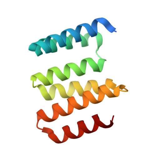
Structure of the Peptidoglycan Synthase Activator LpoP in Pseudomonas aeruginosa.

Caveney, N.A.Egan, A.J.F.Ayala, I.Laguri, C.Robb, C.S.Breukink, E.Vollmer, W.Strynadka, N.C.J.Simorre, J.P.

(2020) Structure 28: 643-650.e5

  • DOI: https://doi.org/10.1016/j.str.2020.03.012
  • Primary Citation of Related Structures:  
    6W5Q

  • PubMed Abstract: 

    Peptidoglycan (PG) is an essential component of the bacterial cell wall and is assembled from a lipid II precursor by glycosyltransferase and transpeptidase reactions catalyzed in particular by bifunctional class A penicillin-binding proteins (aPBPs). In the major clinical pathogen Pseudomonas aeruginosa, PBP1B is anchored within the cytoplasmic membrane but regulated by a bespoke outer membrane-localized lipoprotein known as LpoP. Here, we report the structure of LpoP, showing an extended N-terminal, flexible tether followed by a well-ordered C-terminal tandem-tetratricopeptide repeat domain. We show that LpoP stimulates both PBP1B transpeptidase and glycosyltransferase activities in vitro and interacts directly via its C terminus globular domain with the central UB2H domain of PBP1B. Contrary to the situation in E. coli, P. aeruginosa CpoB does not regulate PBP1B/LpoP in vitro. We propose a mechanism that helps to underscore similarities and differences in class A PBP activation across Gram-negative bacteria.


  • Organizational Affiliation

    Department of Biochemistry and Molecular Biology and Centre for Blood Research, The University of British Columbia, Vancouver V6T 1Z3 BC, Canada.


Macromolecules
Find similar proteins by:  (by identity cutoff)  |  3D Structure
Entity ID: 1
MoleculeChains Sequence LengthOrganismDetailsImage
Peptidoglycan synthase activator LpoP
A, B, C, D, E
A, B, C, D, E, F, G, H
116Pseudomonas aeruginosaMutation(s): 0 
Gene Names: PA4699
UniProt
Find proteins for Q9HV97 (Pseudomonas aeruginosa (strain ATCC 15692 / DSM 22644 / CIP 104116 / JCM 14847 / LMG 12228 / 1C / PRS 101 / PAO1))
Explore Q9HV97 
Go to UniProtKB:  Q9HV97
Entity Groups  
Sequence Clusters30% Identity50% Identity70% Identity90% Identity95% Identity100% Identity
UniProt GroupQ9HV97
Sequence Annotations
Expand
  • Reference Sequence
Small Molecules
Ligands 2 Unique
IDChains Name / Formula / InChI Key2D Diagram3D Interactions
PGE
Query on PGE

Download Ideal Coordinates CCD File 
P [auth E]TRIETHYLENE GLYCOL
C6 H14 O4
ZIBGPFATKBEMQZ-UHFFFAOYSA-N
SO4
Query on SO4

Download Ideal Coordinates CCD File 
I [auth A]
J [auth A]
K [auth B]
L [auth B]
M [auth C]
I [auth A],
J [auth A],
K [auth B],
L [auth B],
M [auth C],
N [auth C],
O [auth D],
Q [auth F],
R [auth F],
S [auth G],
T [auth H],
U [auth H],
V [auth H]
SULFATE ION
O4 S
QAOWNCQODCNURD-UHFFFAOYSA-L
Experimental Data & Validation

Experimental Data

  • Method: X-RAY DIFFRACTION
  • Resolution: 2.20 Å
  • R-Value Free: 0.230 
  • R-Value Work: 0.194 
  • R-Value Observed: 0.196 
  • Space Group: P 1 21 1
Unit Cell:
Length ( Å )Angle ( ˚ )
a = 48.757α = 90
b = 154.584β = 90.15
c = 54.083γ = 90
Software Package:
Software NamePurpose
Aimlessdata scaling
PHENIXrefinement
PDB_EXTRACTdata extraction
XDSdata reduction
PHASERphasing

Structure Validation

View Full Validation Report


Entry History & Funding Information

Deposition Data


Funding OrganizationLocationGrant Number
Canadian Institutes of Health Research (CIHR)Canada--

Revision History  (Full details and data files)

  • Version 1.0: 2020-05-06
    Type: Initial release
  • Version 1.1: 2020-06-10
    Changes: Database references
  • Version 1.2: 2023-10-18
    Changes: Data collection, Database references, Derived calculations, Refinement description