6VLO

X-ray Structure of the R141 Sugar 4,6-dehydratase from Acanthamoeba polyphaga Minivirus


Experimental Data Snapshot

  • Method: X-RAY DIFFRACTION
  • Resolution: 2.05 Å
  • R-Value Free: 0.229 
  • R-Value Work: 0.180 
  • R-Value Observed: 0.183 

wwPDB Validation   3D Report Full Report

Currently 6VLO does not have a validation slider image.


This is version 1.2 of the entry. See complete history


Literature

Biochemical analysis of a sugar 4,6-dehydratase from Acanthamoeba polyphaga Mimivirus.

Ferek, J.D.Thoden, J.B.Holden, H.M.

(2020) Protein Sci 29: 1148-1159

  • DOI: https://doi.org/10.1002/pro.3843
  • Primary Citation of Related Structures:  
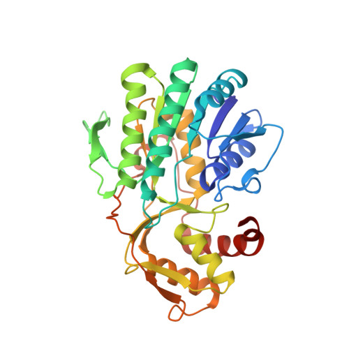
    6VLO

  • PubMed Abstract: 

    The exciting discovery of the giant DNA Mimivirus in 2003 challenged the conventional description of viruses in a radical way, and since then, dozens of additional giant viruses have been identified. It has now been demonstrated that the Mimivirus genome encodes for the two enzymes required for the production of the unusual sugar 4-amino-4,6-dideoxy-d-glucose, namely a 4,6-dehydratase and an aminotransferase. In light of our long-standing interest in the bacterial 4,6-dehydratases and in unusual sugars in general, we conducted a combined structural and functional analysis of the Mimivirus 4,6-dehydratase referred to as R141. For this investigation, the three-dimensional X-ray structure of R141 was determined to 2.05 Å resolution and refined to an R-factor of 18.3%. The overall fold of R141 places it into the short-chain dehydrogenase/reductase (SDR) superfamily of proteins. Whereas its molecular architecture is similar to that observed for the bacterial 4,6-dehydratases, there are two key regions where the polypeptide chain adopts different conformations. In particular, the conserved tyrosine that has been implicated as a catalytic acid or base in SDR superfamily members is splayed away from the active site by nearly 12 Å, thereby suggesting that a major conformational change must occur upon substrate binding. In addition to the structural analysis, the kinetic parameters for R141 using either dTDP-d-glucose or UDP-d-glucose as substrates were determined. Contrary to a previous report, R141 demonstrates nearly identical catalytic efficiency with either nucleotide-linked sugar. The data presented herein represent the first three-dimensional model for a viral 4,6-dehydratase and thus expands our understanding of these fascinating enzymes.


  • Organizational Affiliation

    Department of Biochemistry, University of Wisconsin, Madison, Wisconsin, United States.


Macromolecules
Find similar proteins by:  (by identity cutoff)  |  3D Structure
Entity ID: 1
MoleculeChains Sequence LengthOrganismDetailsImage
Putative dTDP-D-glucose 4,6-dehydratase
A, B, C, D
326Acanthamoeba polyphaga mimivirusMutation(s): 0 
Gene Names: MIMI_R141
EC: 4.2.1.46
UniProt
Find proteins for Q5UR12 (Acanthamoeba polyphaga mimivirus)
Explore Q5UR12 
Go to UniProtKB:  Q5UR12
Entity Groups  
Sequence Clusters30% Identity50% Identity70% Identity90% Identity95% Identity100% Identity
UniProt GroupQ5UR12
Sequence Annotations
Expand
  • Reference Sequence
Small Molecules
Ligands 3 Unique
IDChains Name / Formula / InChI Key2D Diagram3D Interactions
NAD (Subject of Investigation/LOI)
Query on NAD

Download Ideal Coordinates CCD File 
F [auth A],
H [auth B],
K [auth C],
N [auth D]
NICOTINAMIDE-ADENINE-DINUCLEOTIDE
C21 H27 N7 O14 P2
BAWFJGJZGIEFAR-NNYOXOHSSA-N
TYD (Subject of Investigation/LOI)
Query on TYD

Download Ideal Coordinates CCD File 
E [auth A],
G [auth B],
J [auth C],
M [auth D]
THYMIDINE-5'-DIPHOSPHATE
C10 H16 N2 O11 P2
UJLXYODCHAELLY-XLPZGREQSA-N
NI
Query on NI

Download Ideal Coordinates CCD File 
I [auth B],
L [auth C]
NICKEL (II) ION
Ni
VEQPNABPJHWNSG-UHFFFAOYSA-N
Experimental Data & Validation

Experimental Data

  • Method: X-RAY DIFFRACTION
  • Resolution: 2.05 Å
  • R-Value Free: 0.229 
  • R-Value Work: 0.180 
  • R-Value Observed: 0.183 
  • Space Group: P 21 21 21
Unit Cell:
Length ( Å )Angle ( ˚ )
a = 93.883α = 90
b = 111.083β = 90
c = 133.385γ = 90
Software Package:
Software NamePurpose
REFMACrefinement
PDB_EXTRACTdata extraction
HKL-3000data reduction
HKL-3000data scaling
PHASERphasing

Structure Validation

View Full Validation Report

Currently 6VLO does not have a validation slider image.


Entry History & Funding Information

Deposition Data


Funding OrganizationLocationGrant Number
National Institutes of Health/National Institute of General Medical Sciences (NIH/NIGMS)United StatesGM115921

Revision History  (Full details and data files)

  • Version 1.0: 2020-03-04
    Type: Initial release
  • Version 1.1: 2020-05-13
    Changes: Database references
  • Version 1.2: 2023-10-11
    Changes: Data collection, Database references, Derived calculations, Refinement description