6VK7

Crystal Structure of reduced Methylosinus trichosporium OB3b Soluble Methane Monooxygenase Hydroxylase


Experimental Data Snapshot

  • Method: X-RAY DIFFRACTION
  • Resolution: 2.12 Å
  • R-Value Free: 0.203 
  • R-Value Work: 0.162 
  • R-Value Observed: 0.164 

wwPDB Validation   3D Report Full Report

Currently 6VK7 does not have a validation slider image.


This is version 1.3 of the entry. See complete history


Literature

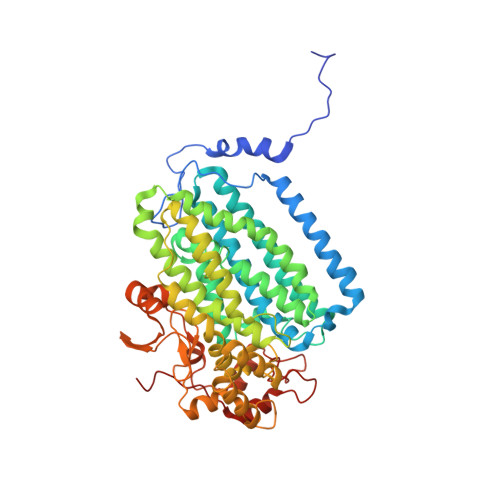
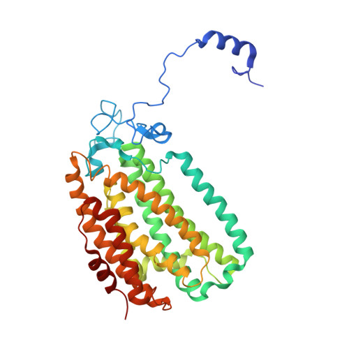
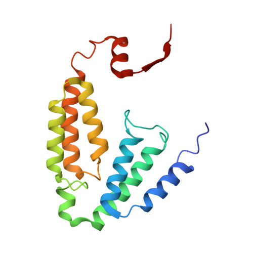
Structural Studies of theMethylosinus trichosporiumOB3b Soluble Methane Monooxygenase Hydroxylase and Regulatory Component Complex Reveal a Transient Substrate Tunnel.

Jones, J.C.Banerjee, R.Shi, K.Aihara, H.Lipscomb, J.D.

(2020) Biochemistry 59: 2946-2961

  • DOI: https://doi.org/10.1021/acs.biochem.0c00459
  • Primary Citation of Related Structures:  
    6VK4, 6VK5, 6VK6, 6VK7, 6VK8

  • PubMed Abstract: 

    The metalloenzyme soluble methane monooxygenase (sMMO) consists of hydroxylase (sMMOH), regulatory (MMOB), and reductase components. When sMMOH forms a complex with MMOB, the rate constants are greatly increased for the sequential access of O 2 , protons, and CH 4 to an oxygen-bridged diferrous metal cluster located in the buried active site. Here, we report high-resolution X-ray crystal structures of the diferric and diferrous states of both sMMOH and the sMMOH:MMOB complex using the components from Methylosinus trichosporium OB3b. These structures are analyzed for O 2 access routes enhanced when the complex forms. Previously reported, lower-resolution structures of the sMMOH:MMOB complex from the sMMO of Methylococcus capsulatus Bath revealed a series of cavities through sMMOH postulated to serve as the O 2 conduit. This potential role is evaluated in greater detail using the current structures. Additionally, a search for other potential O 2 conduits in the M. trichosporium OB3b sMMOH:MMOB complex revealed a narrow molecular tunnel, termed the W308-tunnel. This tunnel is sized appropriately for O 2 and traverses the sMMOH-MMOB interface before accessing the active site. The kinetics of reaction of O 2 with the diferrous sMMOH:MMOB complex in solution show that use of the MMOB V41R variant decreases the rate constant for O 2 binding >25000-fold without altering the component affinity. The location of Val41 near the entrance to the W308-tunnel is consistent with the tunnel serving as the primary route for the transfer of O 2 into the active site. Accordingly, the crystal structures show that formation of the diferrous sMMOH:MMOB complex restricts access through the chain of cavities while opening the W308-tunnel.


Macromolecules
Find similar proteins by:  (by identity cutoff)  |  3D Structure
Entity ID: 1
MoleculeChains Sequence LengthOrganismDetailsImage
Methane monooxygenase component A alpha chain526Methylosinus trichosporium OB3bMutation(s): 0 
UniProt
Find proteins for P27353 (Methylosinus trichosporium)
Explore P27353 
Go to UniProtKB:  P27353
Entity Groups  
Sequence Clusters30% Identity50% Identity70% Identity90% Identity95% Identity100% Identity
UniProt GroupP27353
Sequence Annotations
Expand
  • Reference Sequence
Find similar proteins by:  (by identity cutoff)  |  3D Structure
Entity ID: 2
MoleculeChains Sequence LengthOrganismDetailsImage
Methane monooxygenase395Methylosinus trichosporium OB3bMutation(s): 0 
UniProt
Find proteins for A0A2D2D5X7 (Methylosinus trichosporium OB3b)
Explore A0A2D2D5X7 
Go to UniProtKB:  A0A2D2D5X7
Entity Groups  
Sequence Clusters30% Identity50% Identity70% Identity90% Identity95% Identity100% Identity
UniProt GroupA0A2D2D5X7
Sequence Annotations
Expand
  • Reference Sequence
Find similar proteins by:  (by identity cutoff)  |  3D Structure
Entity ID: 3
MoleculeChains Sequence LengthOrganismDetailsImage
Methane monooxygenase169Methylosinus trichosporium OB3bMutation(s): 0 
UniProt
Find proteins for A0A2D2D0T0 (Methylosinus trichosporium OB3b)
Explore A0A2D2D0T0 
Go to UniProtKB:  A0A2D2D0T0
Entity Groups  
Sequence Clusters30% Identity50% Identity70% Identity90% Identity95% Identity100% Identity
UniProt GroupA0A2D2D0T0
Sequence Annotations
Expand
  • Reference Sequence
Experimental Data & Validation

Experimental Data

  • Method: X-RAY DIFFRACTION
  • Resolution: 2.12 Å
  • R-Value Free: 0.203 
  • R-Value Work: 0.162 
  • R-Value Observed: 0.164 
  • Space Group: C 2 2 21
Unit Cell:
Length ( Å )Angle ( ˚ )
a = 62.458α = 90
b = 290.452β = 90
c = 139.22γ = 90
Software Package:
Software NamePurpose
PHENIXrefinement
XDSdata reduction
PHENIXphasing
PHENIXdata processing
PHENIXdata scaling

Structure Validation

View Full Validation Report

Currently 6VK7 does not have a validation slider image.


Entry History & Funding Information

Deposition Data


Funding OrganizationLocationGrant Number
National Institutes of Health/National Institute of General Medical Sciences (NIH/NIGMS)United StatesGM118030, GM08347
National Institutes of Health/National Institute of General Medical Sciences (NIH/NIGMS)United StatesGM118000, GM118047

Revision History  (Full details and data files)

  • Version 1.0: 2020-08-05
    Type: Initial release
  • Version 1.1: 2020-08-26
    Changes: Database references
  • Version 1.2: 2020-09-02
    Changes: Database references
  • Version 1.3: 2023-10-11
    Changes: Data collection, Database references, Refinement description