6VC0

Crystal structure of the horse MLKL pseudokinase domain


Experimental Data Snapshot

  • Method: X-RAY DIFFRACTION
  • Resolution: 2.75 Å
  • R-Value Free: 0.238 
  • R-Value Work: 0.171 
  • R-Value Observed: 0.175 

wwPDB Validation   3D Report Full Report

Currently 6VC0 does not have a validation slider image.


This is version 1.1 of the entry. See complete history


Literature

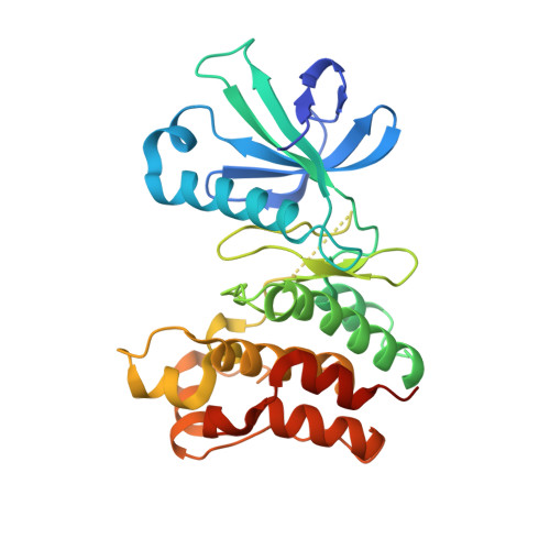
Distinct pseudokinase domain conformations underlie divergent activation mechanisms among vertebrate MLKL orthologues.

Davies, K.A.Fitzgibbon, C.Young, S.N.Garnish, S.E.Yeung, W.Coursier, D.Birkinshaw, R.W.Sandow, J.J.Lehmann, W.I.L.Liang, L.Y.Lucet, I.S.Chalmers, J.D.Patrick, W.M.Kannan, N.Petrie, E.J.Czabotar, P.E.Murphy, J.M.

(2020) Nat Commun 11: 3060-3060

  • DOI: https://doi.org/10.1038/s41467-020-16823-3
  • Primary Citation of Related Structures:  
    6VBZ, 6VC0

  • PubMed Abstract: 

    The MLKL pseudokinase is the terminal effector in the necroptosis cell death pathway. Phosphorylation by its upstream regulator, RIPK3, triggers MLKL's conversion from a dormant cytoplasmic protein into oligomers that translocate to, and permeabilize, the plasma membrane to kill cells. The precise mechanisms underlying these processes are incompletely understood, and were proposed to differ between mouse and human cells. Here, we examine the divergence of activation mechanisms among nine vertebrate MLKL orthologues, revealing remarkable specificity of mouse and human RIPK3 for MLKL orthologues. Pig MLKL can restore necroptotic signaling in human cells; while horse and pig, but not rat, MLKL can reconstitute the mouse pathway. This selectivity can be rationalized from the distinct conformations observed in the crystal structures of horse and rat MLKL pseudokinase domains. These studies identify important differences in necroptotic signaling between species, and suggest that, more broadly, divergent regulatory mechanisms may exist among orthologous pseudoenzymes.


  • Organizational Affiliation

    Walter and Eliza Hall Institute of Medical Research, 1G Royal Parade, Parkville, VIC, 3052, Australia.


Macromolecules
Find similar proteins by:  (by identity cutoff)  |  3D Structure
Entity ID: 1
MoleculeChains Sequence LengthOrganismDetailsImage
Mixed lineage kinase domain like pseudokinase
A, B, C
292Equus caballusMutation(s): 0 
Gene Names: MLKL
UniProt
Find proteins for F6S337 (Equus caballus)
Explore F6S337 
Go to UniProtKB:  F6S337
Entity Groups  
Sequence Clusters30% Identity50% Identity70% Identity90% Identity95% Identity100% Identity
UniProt GroupF6S337
Sequence Annotations
Expand
  • Reference Sequence
Experimental Data & Validation

Experimental Data

  • Method: X-RAY DIFFRACTION
  • Resolution: 2.75 Å
  • R-Value Free: 0.238 
  • R-Value Work: 0.171 
  • R-Value Observed: 0.175 
  • Space Group: C 1 2 1
Unit Cell:
Length ( Å )Angle ( ˚ )
a = 155.532α = 90
b = 124.775β = 112.75
c = 81.053γ = 90
Software Package:
Software NamePurpose
PHENIXrefinement
XSCALEdata scaling
PDB_EXTRACTdata extraction
XDSdata reduction
PHASERphasing

Structure Validation

View Full Validation Report

Currently 6VC0 does not have a validation slider image.


Entry History & Funding Information

Deposition Data


Funding OrganizationLocationGrant Number
National Health and Medical Research Council (NHMRC, Australia)--

Revision History  (Full details and data files)

  • Version 1.0: 2020-07-08
    Type: Initial release
  • Version 1.1: 2023-10-11
    Changes: Data collection, Database references, Refinement description