6UVK

OXA-48 bound by inhibitor CDD-97


Experimental Data Snapshot

  • Method: X-RAY DIFFRACTION
  • Resolution: 2.20 Å
  • R-Value Free: 0.223 
  • R-Value Work: 0.184 
  • R-Value Observed: 0.186 

wwPDB Validation   3D Report Full Report


Ligand Structure Quality Assessment 


This is version 1.4 of the entry. See complete history


Literature

Identifying Oxacillinase-48 Carbapenemase Inhibitors Using DNA-Encoded Chemical Libraries.

Taylor, D.M.Anglin, J.Park, S.Ucisik, M.N.Faver, J.C.Simmons, N.Jin, Z.Palaniappan, M.Nyshadham, P.Li, F.Campbell, J.Hu, L.Sankaran, B.Prasad, B.V.V.Huang, H.Matzuk, M.M.Palzkill, T.

(2020) ACS Infect Dis 6: 1214-1227

  • DOI: https://doi.org/10.1021/acsinfecdis.0c00015
  • Primary Citation of Related Structures:  
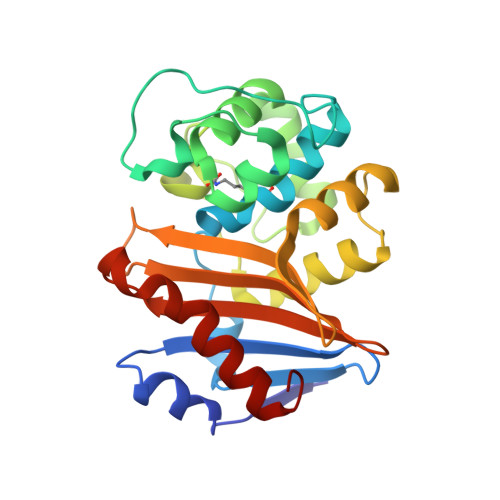
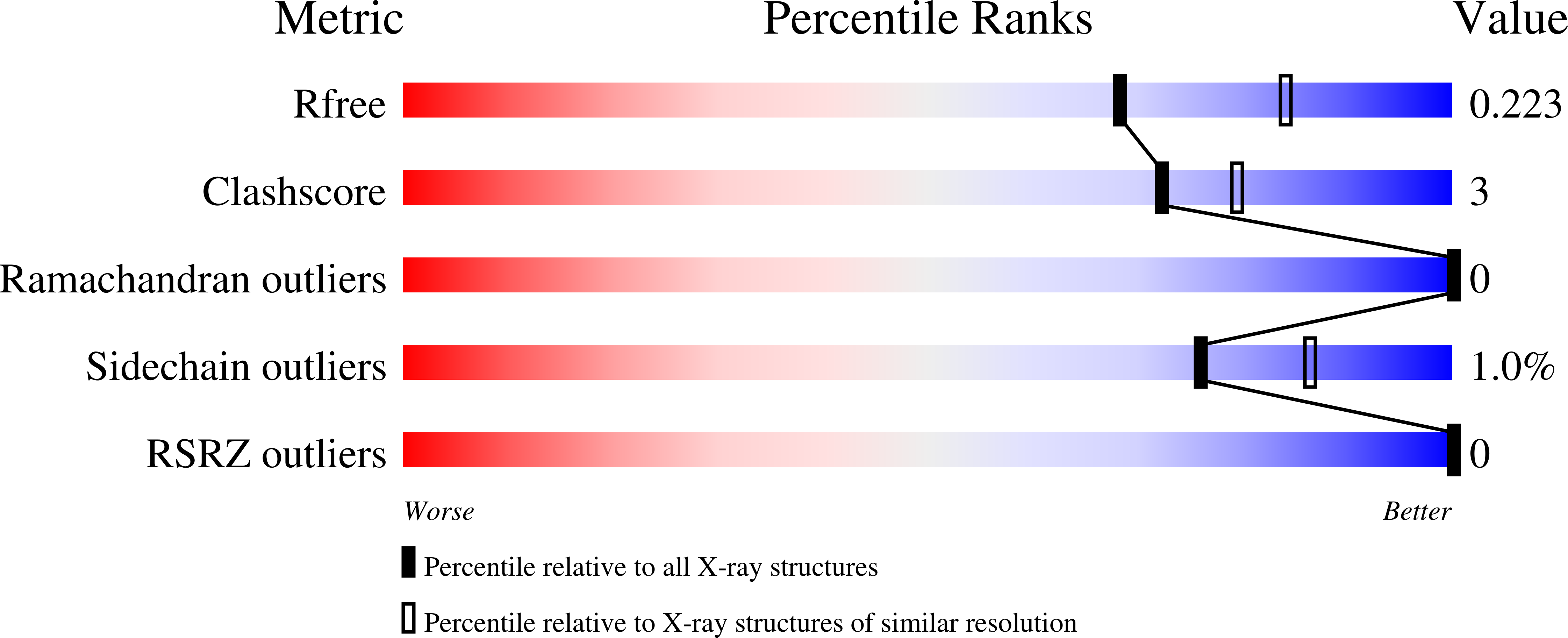
    6UVK

  • PubMed Abstract: 

    Bacterial resistance to β-lactam antibiotics is largely mediated by β-lactamases, which catalyze the hydrolysis of these drugs and continue to emerge in response to antibiotic use. β-Lactamases that hydrolyze the last resort carbapenem class of β-lactam antibiotics (carbapenemases) are a growing global health threat. Inhibitors have been developed to prevent β-lactamase-mediated hydrolysis and restore the efficacy of these antibiotics. However, there are few inhibitors available for problematic carbapenemases such as oxacillinase-48 (OXA-48). A DNA-encoded chemical library approach was used to rapidly screen for compounds that bind and potentially inhibit OXA-48. Using this approach, a hit compound, CDD-97, was identified with submicromolar potency ( K i = 0.53 ± 0.08 μM) against OXA-48. X-ray crystallography showed that CDD-97 binds noncovalently in the active site of OXA-48. Synthesis and testing of derivatives of CDD-97 revealed structure-activity relationships and informed the design of a compound with a 2-fold increase in potency. CDD-97, however, synergizes poorly with β-lactam antibiotics to inhibit the growth of bacteria expressing OXA-48 due to poor accumulation into E. coli . Despite the low in vivo activity, CDD-97 provides new insights into OXA-48 inhibition and demonstrates the potential of using DNA-encoded chemistry technology to rapidly identify β-lactamase binders and to study β-lactamase inhibition, leading to clinically useful inhibitors.


  • Organizational Affiliation

    Verna and Marrs McLean Department of Biochemistry and Molecular Biology, Baylor College of Medicine, Houston, Texas 77030, United States.


Macromolecules
Find similar proteins by:  (by identity cutoff)  |  3D Structure
Entity ID: 1
MoleculeChains Sequence LengthOrganismDetailsImage
Beta-lactamase
A, B, C, D
244Klebsiella pneumoniaeMutation(s): 0 
Gene Names: bla OXA-48blaOXA-48FNY87_29090KPE71T_00045
EC: 3.5.2.6
UniProt
Find proteins for Q6XEC0 (Klebsiella pneumoniae)
Explore Q6XEC0 
Go to UniProtKB:  Q6XEC0
Entity Groups  
Sequence Clusters30% Identity50% Identity70% Identity90% Identity95% Identity100% Identity
UniProt GroupQ6XEC0
Sequence Annotations
Expand
  • Reference Sequence
Small Molecules
Ligands 4 Unique
IDChains Name / Formula / InChI Key2D Diagram3D Interactions
QHY (Subject of Investigation/LOI)
Query on QHY

Download Ideal Coordinates CCD File 
E [auth A],
G [auth B],
I [auth C],
L [auth D]
1-{4-[4-(2-ethoxyphenyl)piperazin-1-yl]-1,3,5-triazin-2-yl}piperidine-4-carboxylic acid
C21 H28 N6 O3
MFDIQEKYAZCUMU-UHFFFAOYSA-N
GOL
Query on GOL

Download Ideal Coordinates CCD File 
J [auth C]GLYCEROL
C3 H8 O3
PEDCQBHIVMGVHV-UHFFFAOYSA-N
EDO
Query on EDO

Download Ideal Coordinates CCD File 
F [auth A],
H [auth B]
1,2-ETHANEDIOL
C2 H6 O2
LYCAIKOWRPUZTN-UHFFFAOYSA-N
CL
Query on CL

Download Ideal Coordinates CCD File 
K [auth C],
M [auth D],
N [auth D]
CHLORIDE ION
Cl
VEXZGXHMUGYJMC-UHFFFAOYSA-M
Modified Residues  1 Unique
IDChains TypeFormula2D DiagramParent
KCX
Query on KCX
A, B, C, D
L-PEPTIDE LINKINGC7 H14 N2 O4LYS
Binding Affinity Annotations 
IDSourceBinding Affinity
QHY Binding MOAD:  6UVK Ki: 530 (nM) from 1 assay(s)
Experimental Data & Validation

Experimental Data

  • Method: X-RAY DIFFRACTION
  • Resolution: 2.20 Å
  • R-Value Free: 0.223 
  • R-Value Work: 0.184 
  • R-Value Observed: 0.186 
  • Space Group: P 65
Unit Cell:
Length ( Å )Angle ( ˚ )
a = 123.054α = 90
b = 123.054β = 90
c = 161.329γ = 120
Software Package:
Software NamePurpose
PHENIXrefinement
HKL-2000data reduction
HKL-2000data scaling
PHASERphasing

Structure Validation

View Full Validation Report


Ligand Structure Quality Assessment 


Entry History & Funding Information

Deposition Data


Funding OrganizationLocationGrant Number
National Institutes of Health/National Institute Of Allergy and Infectious Diseases (NIH/NIAID)United StatesAI32956
Department of Energy (DOE, United States)United StatesDE-AC02-05CH11231
National Institutes of Health/National Institute of General Medical Sciences (NIH/NIGMS)United StatesT32GM120011

Revision History  (Full details and data files)

  • Version 1.0: 2020-05-06
    Type: Initial release
  • Version 1.1: 2020-05-20
    Changes: Database references
  • Version 1.2: 2021-06-30
    Changes: Structure summary
  • Version 1.3: 2023-10-11
    Changes: Data collection, Database references, Refinement description
  • Version 1.4: 2023-11-15
    Changes: Data collection