6U3A

1.65 Angstrom crystal structure of the N97S Ca-CaM:CaV1.2 IQ domain complex


Experimental Data Snapshot

  • Method: X-RAY DIFFRACTION
  • Resolution: 1.65 Å
  • R-Value Free: 0.209 
  • R-Value Work: 0.178 
  • R-Value Observed: 0.179 

wwPDB Validation   3D Report Full Report


This is version 1.2 of the entry. See complete history


Literature

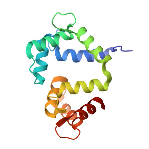
Arrhythmia mutations in calmodulin can disrupt cooperativity of Ca2+binding and cause misfolding.

Wang, K.Brohus, M.Holt, C.Overgaard, M.T.Wimmer, R.Van Petegem, F.

(2020) J Physiol 598: 1169-1186

  • DOI: https://doi.org/10.1113/JP279307
  • Primary Citation of Related Structures:  
    6U39, 6U3A, 6U3B, 6U3D

  • PubMed Abstract: 

    Mutations in the calmodulin protein (CaM) are associated with arrhythmia syndromes. This study focuses on understanding the structural characteristics of CaM disease mutants and their interactions with the voltage-gated calcium channel Ca V 1.2. Arrhythmia mutations in CaM can lead to loss of Ca 2+ binding, uncoupling of Ca 2+ binding cooperativity, misfolding of the EF-hands and altered affinity for the calcium channel. These results help us to understand how different CaM mutants have distinct effects on structure and interactions with protein targets to cause disease. Calmodulinopathies are life-threatening arrhythmia syndromes that arise from mutations in calmodulin (CaM), a calcium sensing protein whose sequence is completely conserved across all vertebrates. These mutations have been shown to interfere with the function of cardiac ion channels, including the voltage-gated Ca 2+ channel Ca V 1.2 and the ryanodine receptor (RyR2), in a mutation-specific manner. The ability of different CaM disease mutations to discriminate between these channels has been enigmatic. We present crystal structures of several C-terminal lobe mutants and an N-terminal lobe mutant in complex with the Ca V 1.2 IQ domain, in conjunction with binding assays and complementary structural biology techniques. One mutation (D130G) causes a pathological conformation, with complete separation of EF-hands within the C-lobe and loss of Ca 2+ binding in EF-hand 4. Another variant (Q136P) has severely reduced affinity for the IQ domain, and shows changes in the CD spectra under Ca 2+ -saturating conditions when unbound to the IQ domain. Ca 2+ binding to a pair of EF-hands normally proceeds with very high cooperativity, but we found that N98S CaM can adopt different conformations with either one or two Ca 2+ ions bound to the C-lobe, possibly disrupting the cooperativity. An N-lobe variant (N54I), which causes severe stress-induced arrhythmia, does not show any major changes in complex with the IQ domain, providing a structural basis for why this mutant does not affect function of Ca V 1.2. These findings show that different CaM mutants have distinct effects on both the CaM structure and interactions with protein targets, and act via distinct pathological mechanisms to cause disease.


  • Organizational Affiliation

    Department of Biochemistry and Molecular Biology, Life Sciences Institute, University of British Columbia, V6T 1Z3 Vancouver, BC, Canada.


Macromolecules
Find similar proteins by:  (by identity cutoff)  |  3D Structure
Entity ID: 1
MoleculeChains Sequence LengthOrganismDetailsImage
Calmodulin-1A,
C [auth B]
148Homo sapiensMutation(s): 1 
Gene Names: CALM1CALMCAMCAM1
UniProt & NIH Common Fund Data Resources
Find proteins for P0DP23 (Homo sapiens)
Explore P0DP23 
Go to UniProtKB:  P0DP23
PHAROS:  P0DP23
GTEx:  ENSG00000198668 
Entity Groups  
Sequence Clusters30% Identity50% Identity70% Identity90% Identity95% Identity100% Identity
UniProt GroupP0DP23
Sequence Annotations
Expand
  • Reference Sequence
Find similar proteins by:  (by identity cutoff)  |  3D Structure
Entity ID: 2
MoleculeChains Sequence LengthOrganismDetailsImage
Voltage-dependent L-type calcium channel subunit alpha-1CB [auth C],
D
37Homo sapiensMutation(s): 0 
Gene Names: CACNA1CCACH2CACN2CACNL1A1CCHL1A1
UniProt & NIH Common Fund Data Resources
Find proteins for Q13936 (Homo sapiens)
Explore Q13936 
Go to UniProtKB:  Q13936
PHAROS:  Q13936
GTEx:  ENSG00000151067 
Entity Groups  
Sequence Clusters30% Identity50% Identity70% Identity90% Identity95% Identity100% Identity
UniProt GroupQ13936
Sequence Annotations
Expand
  • Reference Sequence
Small Molecules
Ligands 2 Unique
IDChains Name / Formula / InChI Key2D Diagram3D Interactions
CA (Subject of Investigation/LOI)
Query on CA

Download Ideal Coordinates CCD File 
E [auth A]
F [auth A]
G [auth A]
J [auth B]
K [auth B]
E [auth A],
F [auth A],
G [auth A],
J [auth B],
K [auth B],
L [auth B],
M [auth B]
CALCIUM ION
Ca
BHPQYMZQTOCNFJ-UHFFFAOYSA-N
NA (Subject of Investigation/LOI)
Query on NA

Download Ideal Coordinates CCD File 
H [auth A],
I [auth A]
SODIUM ION
Na
FKNQFGJONOIPTF-UHFFFAOYSA-N
Experimental Data & Validation

Experimental Data

  • Method: X-RAY DIFFRACTION
  • Resolution: 1.65 Å
  • R-Value Free: 0.209 
  • R-Value Work: 0.178 
  • R-Value Observed: 0.179 
  • Space Group: P 21 21 21
Unit Cell:
Length ( Å )Angle ( ˚ )
a = 63.419α = 90
b = 68.077β = 90
c = 86.25γ = 90
Software Package:
Software NamePurpose
XSCALEdata scaling
PHENIXrefinement
PDB_EXTRACTdata extraction
XDSdata reduction
PHASERphasing

Structure Validation

View Full Validation Report


Entry History & Funding Information

Deposition Data


Funding OrganizationLocationGrant Number
Canadian Institutes of Health Research (CIHR)Canada--

Revision History  (Full details and data files)

  • Version 1.0: 2020-02-19
    Type: Initial release
  • Version 1.1: 2020-03-25
    Changes: Database references
  • Version 1.2: 2023-10-11
    Changes: Data collection, Database references, Derived calculations, Refinement description