6TLD

Crystal structure of Schistosoma mansoni HDAC8 complexed with a triazole hydroxamate inhibitor 2


Experimental Data Snapshot

  • Method: X-RAY DIFFRACTION
  • Resolution: 1.61 Å
  • R-Value Free: 0.195 
  • R-Value Work: 0.166 
  • R-Value Observed: 0.167 

wwPDB Validation   3D Report Full Report


Ligand Structure Quality Assessment 


This is version 1.2 of the entry. See complete history


Literature

Structure-Based Design, Synthesis, and Biological Evaluation of Triazole-Based smHDAC8 Inhibitors.

Kalinin, D.V.Jana, S.K.Pfafenrot, M.Chakrabarti, A.Melesina, J.Shaik, T.B.Lancelot, J.Pierce, R.J.Sippl, W.Romier, C.Jung, M.Holl, R.

(2020) ChemMedChem 15: 571-584

  • DOI: https://doi.org/10.1002/cmdc.201900583
  • Primary Citation of Related Structures:  
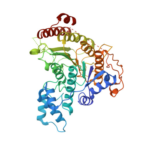
    6TLD

  • PubMed Abstract: 

    Schistosomiasis is a neglected tropical disease caused by parasitic flatworms of the genus Schistosoma, which affects over 200 million people worldwide and leads to at least 300,000 deaths every year. In this study, initial screening revealed the triazole-based hydroxamate 2 b (N-hydroxy-1-phenyl-1H-1,2,3-triazole-4-carboxamide) exhibiting potent inhibitory activity toward the novel antiparasitic target Schistosoma mansoni histone deacetylase 8 (smHDAC8) and promising selectivity over the major human HDACs. Subsequent crystallographic studies of the 2 b/smHDAC8 complex revealed key interactions between the inhibitor and the enzyme's active site, thus explaining the unique selectivity profile of the inhibitor. Further chemical modifications of 2 b led to the discovery of 4-fluorophenoxy derivative 21 (1-[5-chloro-2-(4-fluorophenoxy)phenyl]-N-hydroxy-1H-1,2,3-triazole-4-carboxamide), a nanomolar smHDAC8 inhibitor (IC 50 =0.5 μM), exceeding the smHDAC8 inhibitory activity of 2 b and SAHA (vorinostat), while exhibiting an improved selectivity profile over the investigated human HDACs. Collectively, this study reveals specific interactions between smHDAC8 and the synthesized triazole-based inhibitors and demonstrates that these small molecules represent promising lead structures, which could be further developed in the search for novel drugs for the treatment of schistosomiasis.


  • Organizational Affiliation

    Department of Chemistry Institute of Organic Chemistry, University of Hamburg, Martin-Luther-King-Platz 6, 20146, Hamburg, Germany.


Macromolecules
Find similar proteins by:  (by identity cutoff)  |  3D Structure
Entity ID: 1
MoleculeChains Sequence LengthOrganismDetailsImage
Histone deacetylase
A, B, C, D
447Schistosoma mansoniMutation(s): 0 
Gene Names: HDAC8
UniProt
Find proteins for A5H660 (Schistosoma mansoni)
Explore A5H660 
Go to UniProtKB:  A5H660
Entity Groups  
Sequence Clusters30% Identity50% Identity70% Identity90% Identity95% Identity100% Identity
UniProt GroupA5H660
Sequence Annotations
Expand
  • Reference Sequence
Small Molecules
Ligands 5 Unique
IDChains Name / Formula / InChI Key2D Diagram3D Interactions
NK5 (Subject of Investigation/LOI)
Query on NK5

Download Ideal Coordinates CCD File 
DA [auth C],
H [auth A],
PA [auth D],
R [auth B]
~{N}-oxidanyl-1-phenyl-1,2,3-triazole-4-carboxamide
C9 H8 N4 O2
ALZYWLPUOFGSEC-UHFFFAOYSA-N
GOL
Query on GOL

Download Ideal Coordinates CCD File 
JA [auth C]
KA [auth C]
L [auth A]
M [auth A]
XA [auth D]
JA [auth C],
KA [auth C],
L [auth A],
M [auth A],
XA [auth D],
Z [auth B]
GLYCEROL
C3 H8 O3
PEDCQBHIVMGVHV-UHFFFAOYSA-N
DMF
Query on DMF

Download Ideal Coordinates CCD File 
EA [auth C]
FA [auth C]
GA [auth C]
HA [auth C]
I [auth A]
EA [auth C],
FA [auth C],
GA [auth C],
HA [auth C],
I [auth A],
IA [auth C],
J [auth A],
K [auth A],
LA [auth D],
N [auth A],
QA [auth D],
RA [auth D],
S [auth B],
SA [auth D],
T [auth B],
TA [auth D],
U [auth B],
UA [auth D],
V [auth B],
VA [auth D],
W [auth B],
WA [auth D],
X [auth B],
Y [auth B]
DIMETHYLFORMAMIDE
C3 H7 N O
ZMXDDKWLCZADIW-UHFFFAOYSA-N
ZN
Query on ZN

Download Ideal Coordinates CCD File 
AA [auth C],
E [auth A],
MA [auth D],
O [auth B]
ZINC ION
Zn
PTFCDOFLOPIGGS-UHFFFAOYSA-N
K
Query on K

Download Ideal Coordinates CCD File 
BA [auth C]
CA [auth C]
F [auth A]
G [auth A]
NA [auth D]
BA [auth C],
CA [auth C],
F [auth A],
G [auth A],
NA [auth D],
OA [auth D],
P [auth B],
Q [auth B]
POTASSIUM ION
K
NPYPAHLBTDXSSS-UHFFFAOYSA-N
Binding Affinity Annotations 
IDSourceBinding Affinity
NK5 Binding MOAD:  6TLD Kd: 4400 (nM) from 1 assay(s)
Experimental Data & Validation

Experimental Data

  • Method: X-RAY DIFFRACTION
  • Resolution: 1.61 Å
  • R-Value Free: 0.195 
  • R-Value Work: 0.166 
  • R-Value Observed: 0.167 
  • Space Group: P 1
Unit Cell:
Length ( Å )Angle ( ˚ )
a = 70.64α = 78.08
b = 70.72β = 75.69
c = 98.05γ = 85.72
Software Package:
Software NamePurpose
PHENIXrefinement
PDB_EXTRACTdata extraction
XDSdata reduction
XDSdata scaling
PHENIXphasing

Structure Validation

View Full Validation Report


Ligand Structure Quality Assessment 


Entry History & Funding Information

Deposition Data


Funding OrganizationLocationGrant Number
European CommissionFrance241865
European CommissionFrance602080

Revision History  (Full details and data files)

  • Version 1.0: 2019-12-18
    Type: Initial release
  • Version 1.1: 2020-04-08
    Changes: Database references
  • Version 1.2: 2024-01-24
    Changes: Data collection, Database references, Derived calculations, Refinement description