Lateral A11type tetramerization in lamins.
Lilina, A.V., Chernyatina, A.A., Guzenko, D., Strelkov, S.V.(2020) J Struct Biol 209: 107404-107404
- PubMed: 31610238
- DOI: https://doi.org/10.1016/j.jsb.2019.10.006
- Primary Citation of Related Structures:
6SNZ - PubMed Abstract:
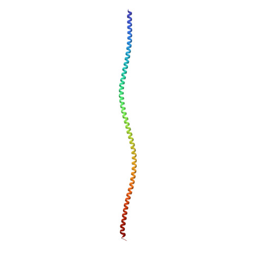
The assembly of intermediate filaments (IFs) including nuclear lamins is driven by specific interactions of the elementary coiled-coil dimers in both lateral and longitudinal direction. The assembly mode A 11 is dependent on lateral tetramerization of the second coiled-coil segment (coil1b) in antiparallel fashion. Recent cryo-electron microscopy studies pointed to 3.5 nm lamin filaments built from two antiparallel threads of longitudinally associated dimers but little molecular detail is available to date. Here we present the 2.6 Å resolution X-ray structure of a lamin A fragment including residues 65-222 which reveals the molecular basis of the A 11 interaction. The crystal structure also indicates a continuous α-helical structure for the preceding linker L1 region. The middle part of the antiparallel tetramer reveals unique interactions due to the lamin-specific 42-residue insert in coil1b. At the same time, distinct characteristics of this insert provide for the preservation of common structural principles shared with lateral coil1b tetramers of vimentin and keratin K1/K10. In addition, structural analysis suggests that the A 11 interaction in lamins is somewhat weaker than in cytoplasmic IFs, despite a 30% longer overlap. Establishing the structural detail of the A 11 interaction across IF types is the first step towards a rational understanding of the IF assembly process which is indispensable for establishing the mechanism of disease-related mutations.
Organizational Affiliation:
Laboratory for Biocrystallography, Department of Pharmaceutical and Pharmacological Sciences, KU Leuven, Belgium.