6SIR

Crystal structure of the guanylate cyclase domain of RhGC from Catenaria anguillulae in complex with GTP


Experimental Data Snapshot

  • Method: X-RAY DIFFRACTION
  • Resolution: 1.70 Å
  • R-Value Free: 0.209 
  • R-Value Work: 0.182 
  • R-Value Observed: 0.183 

wwPDB Validation   3D Report Full Report


Ligand Structure Quality Assessment 


This is version 1.2 of the entry. See complete history


Literature

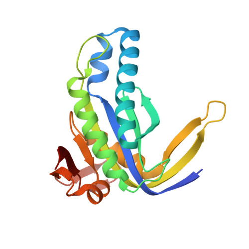
Molecular basis for GTP recognition by light-activated guanylate cyclase RhGC.

Butryn, A.Raza, H.Rada, H.Moraes, I.Owens, R.J.Orville, A.M.

(2020) FEBS J 287: 2797-2807

  • DOI: https://doi.org/10.1111/febs.15167
  • Primary Citation of Related Structures:  
    6SIR

  • PubMed Abstract: 

    Cyclic guanosine 3',5'-monophosphate (cGMP) is an intracellular signalling molecule involved in many sensory and developmental processes. Synthesis of cGMP from GTP is catalysed by guanylate cyclase (GC) in a reaction analogous to cAMP formation by adenylate cyclase (AC). Although detailed structural information is available on the catalytic region of nucleotidyl cyclases (NCs) in various states, these atomic models do not provide a sufficient explanation for the substrate selectivity between GC and AC family members. Detailed structural information on the GC domain in its active conformation is largely missing, and no crystal structure of a GTP-bound wild-type GC domain has been published to date. Here, we describe the crystal structure of the catalytic domain of rhodopsin-GC (RhGC) from Catenaria anguillulae in complex with GTP at 1.7 Å resolution. Our study reveals the organization of a eukaryotic GC domain in its active conformation. We observe that the binding mode of the substrate GTP is similar to that of AC-ATP interaction, although surprisingly not all of the interactions predicted to be responsible for base recognition are present. The structure provides insights into potential mechanisms of substrate discrimination and activity regulation that may be common to all class III purine NCs. DATABASE: Structural data are available in Protein Data Bank database under the accession number 6SIR. ENZYMES: EC4.6.1.2.


  • Organizational Affiliation

    Diamond Light Source Limited, Didcot, UK.


Macromolecules
Find similar proteins by:  (by identity cutoff)  |  3D Structure
Entity ID: 1
MoleculeChains Sequence LengthOrganismDetailsImage
Nucleotide cyclaseA,
B,
C [auth D],
D [auth C]
187Catenaria anguillulae PL171Mutation(s): 0 
Gene Names: BCR44DRAFT_1480233
UniProt
Find proteins for A0A1Y2HEJ3 (Catenaria anguillulae PL171)
Explore A0A1Y2HEJ3 
Go to UniProtKB:  A0A1Y2HEJ3
Entity Groups  
Sequence Clusters30% Identity50% Identity70% Identity90% Identity95% Identity100% Identity
UniProt GroupA0A1Y2HEJ3
Sequence Annotations
Expand
  • Reference Sequence
Small Molecules
Ligands 3 Unique
IDChains Name / Formula / InChI Key2D Diagram3D Interactions
GTP
Query on GTP

Download Ideal Coordinates CCD File 
G [auth A],
I [auth B],
K [auth D],
N [auth C]
GUANOSINE-5'-TRIPHOSPHATE
C10 H16 N5 O14 P3
XKMLYUALXHKNFT-UUOKFMHZSA-N
GOL
Query on GOL

Download Ideal Coordinates CCD File 
F [auth A],
L [auth D]
GLYCEROL
C3 H8 O3
PEDCQBHIVMGVHV-UHFFFAOYSA-N
CA
Query on CA

Download Ideal Coordinates CCD File 
E [auth A],
H [auth B],
J [auth D],
M [auth C]
CALCIUM ION
Ca
BHPQYMZQTOCNFJ-UHFFFAOYSA-N
Experimental Data & Validation

Experimental Data

  • Method: X-RAY DIFFRACTION
  • Resolution: 1.70 Å
  • R-Value Free: 0.209 
  • R-Value Work: 0.182 
  • R-Value Observed: 0.183 
  • Space Group: P 21 21 21
Unit Cell:
Length ( Å )Angle ( ˚ )
a = 58.65α = 90
b = 95.17β = 90
c = 138.66γ = 90
Software Package:
Software NamePurpose
PHENIXrefinement
DIALSdata reduction
Aimlessdata scaling
PHASERphasing

Structure Validation

View Full Validation Report


Ligand Structure Quality Assessment 


Entry History & Funding Information

Deposition Data


Funding OrganizationLocationGrant Number
Wellcome TrustUnited Kingdom102593
Wellcome TrustUnited Kingdom210734/Z/18/Z
Royal SocietyUnited KingdomRSWF/R2/182017

Revision History  (Full details and data files)

  • Version 1.0: 2019-12-18
    Type: Initial release
  • Version 1.1: 2020-07-15
    Changes: Database references
  • Version 1.2: 2024-01-24
    Changes: Data collection, Database references, Derived calculations, Refinement description