6SA0

Ternary complex of Prim-PolC from Mycobacterium smegmatis with 2nt gapped DNA and UpNHpp


Experimental Data Snapshot

  • Method: X-RAY DIFFRACTION
  • Resolution: 2.21 Å
  • R-Value Free: 0.206 
  • R-Value Work: 0.174 
  • R-Value Observed: 0.176 

wwPDB Validation   3D Report Full Report


Ligand Structure Quality Assessment 


This is version 1.1 of the entry. See complete history


Literature

Molecular basis for DNA repair synthesis on short gaps by mycobacterial Primase-Polymerase C.

Brissett, N.C.Zabrady, K.Plocinski, P.Bianchi, J.Korycka-Machala, M.Brzostek, A.Dziadek, J.Doherty, A.J.

(2020) Nat Commun 11: 4196-4196

  • DOI: https://doi.org/10.1038/s41467-020-18012-8
  • Primary Citation of Related Structures:  
    6SA0, 6SA1

  • PubMed Abstract: 

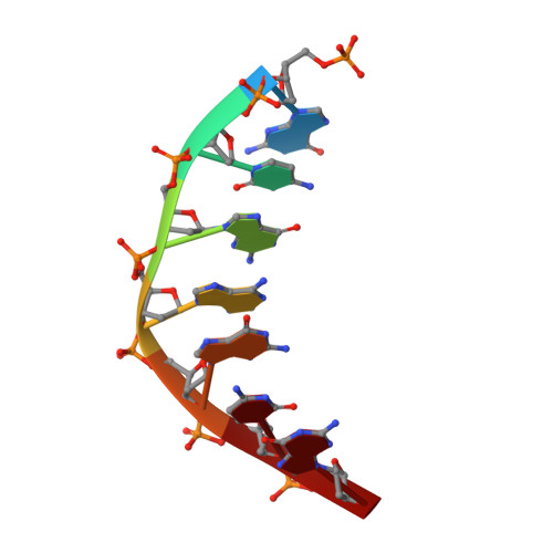
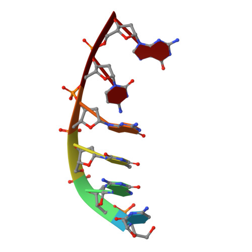
    Cells utilise specialized polymerases from the Primase-Polymerase (Prim-Pol) superfamily to maintain genome stability. Prim-Pol's function in genome maintenance pathways including replication, repair and damage tolerance. Mycobacteria contain multiple Prim-Pols required for lesion repair, including Prim-PolC that performs short gap repair synthesis during excision repair. To understand the molecular basis of Prim-PolC's gap recognition and synthesis activities, we elucidated crystal structures of pre- and post-catalytic complexes bound to gapped DNA substrates. These intermediates explain its binding preference for short gaps and reveal a distinctive modus operandi called Synthesis-dependent Template Displacement (STD). This mechanism enables Prim-PolC to couple primer extension with template base dislocation, ensuring that the unpaired templating bases in the gap are ushered into the active site in an ordered manner. Insights provided by these structures establishes the molecular basis of Prim-PolC's gap recognition and extension activities, while also illuminating the mechanisms of primer extension utilised by closely related Prim-Pols.


  • Organizational Affiliation

    Genome Damage and Stability Centre, School of Life Sciences, University of Sussex, Brighton, BN1 9RQ, UK.


Macromolecules

Find similar proteins by:  (by identity cutoff)  |  3D Structure
Entity ID: 1
MoleculeChains Sequence LengthOrganismDetailsImage
DNA polymerase LigD, polymerase domainA,
E [auth D]
350Mycolicibacterium smegmatis MC2 155Mutation(s): 0 
Gene Names: ligD
UniProt
Find proteins for A0R5T1 (Mycolicibacterium smegmatis (strain ATCC 700084 / mc(2)155))
Explore A0R5T1 
Go to UniProtKB:  A0R5T1
Entity Groups  
Sequence Clusters30% Identity50% Identity70% Identity90% Identity95% Identity100% Identity
UniProt GroupA0R5T1
Sequence Annotations
Expand
  • Reference Sequence

Find similar nucleic acids by:  Sequence   |   3D Structure  

Entity ID: 2
MoleculeChains LengthOrganismImage
DNA (5'-D(P*GP*CP*GP*AP*GP*CP*G)-3')B,
F [auth E]
7synthetic construct
Sequence Annotations
Expand
  • Reference Sequence

Find similar nucleic acids by:  Sequence   |   3D Structure  

Entity ID: 3
MoleculeChains LengthOrganismImage
DNA (5'-D(*CP*GP*CP*TP*CP*GP*CP*AP*AP*CP*GP*CP*A)-3')C,
G [auth F]
15synthetic construct
Sequence Annotations
Expand
  • Reference Sequence

Find similar nucleic acids by:  Sequence   |   3D Structure  

Entity ID: 4
MoleculeChains LengthOrganismImage
DNA (5'-D(P*TP*GP*CP*GP)-3')D [auth H],
H [auth G]
6synthetic construct
Sequence Annotations
Expand
  • Reference Sequence
Small Molecules
Ligands 4 Unique
IDChains Name / Formula / InChI Key2D Diagram3D Interactions
2KH (Subject of Investigation/LOI)
Query on 2KH

Download Ideal Coordinates CCD File 
I [auth A],
O [auth D]
5'-O-[(S)-hydroxy{[(S)-hydroxy(phosphonooxy)phosphoryl]amino}phosphoryl]uridine
C9 H16 N3 O14 P3
OZIBFYOFLVBDIY-XVFCMESISA-N
CAC
Query on CAC

Download Ideal Coordinates CCD File 
L [auth A]CACODYLATE ION
C2 H6 As O2
OGGXGZAMXPVRFZ-UHFFFAOYSA-M
EDO
Query on EDO

Download Ideal Coordinates CCD File 
M [auth A],
N [auth A],
R [auth D]
1,2-ETHANEDIOL
C2 H6 O2
LYCAIKOWRPUZTN-UHFFFAOYSA-N
MN
Query on MN

Download Ideal Coordinates CCD File 
J [auth A],
K [auth A],
P [auth D],
Q [auth D]
MANGANESE (II) ION
Mn
WAEMQWOKJMHJLA-UHFFFAOYSA-N
Experimental Data & Validation

Experimental Data

  • Method: X-RAY DIFFRACTION
  • Resolution: 2.21 Å
  • R-Value Free: 0.206 
  • R-Value Work: 0.174 
  • R-Value Observed: 0.176 
  • Space Group: P 31 2 1
Unit Cell:
Length ( Å )Angle ( ˚ )
a = 136.059α = 90
b = 136.059β = 90
c = 133.479γ = 120
Software Package:
Software NamePurpose
DIALSdata reduction
xia2data scaling
PHASERphasing
PHENIXrefinement
PDB_EXTRACTdata extraction
Cootmodel building

Structure Validation

View Full Validation Report


Ligand Structure Quality Assessment 


Entry History & Funding Information

Deposition Data


Funding OrganizationLocationGrant Number
Biotechnology and Biological Sciences Research CouncilUnited KingdomBB/F013795/1
Biotechnology and Biological Sciences Research CouncilUnited KingdomBB/J018643/1
Biotechnology and Biological Sciences Research CouncilUnited KingdomBB/M004236/1

Revision History  (Full details and data files)

  • Version 1.0: 2020-11-18
    Type: Initial release
  • Version 1.1: 2024-01-24
    Changes: Data collection, Database references, Refinement description