Structural and catalytic characterization of Blastochloris viridis and Pseudomonas aeruginosa homospermidine synthases supports the essential role of cation-pi interaction.
Helfrich, F., Scheidig, A.J.(2021) Acta Crystallogr D Struct Biol 77: 1317-1335
- PubMed: 34605434
- DOI: https://doi.org/10.1107/S2059798321008937
- Primary Citation of Related Structures:
6S3X, 6S49, 6S4D, 6S6G, 6S72, 6SEP, 6Y87 - PubMed Abstract:
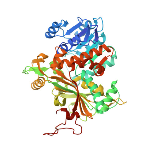
Polyamines influence medically relevant processes in the opportunistic pathogen Pseudomonas aeruginosa, including virulence, biofilm formation and susceptibility to antibiotics. Although homospermidine synthase (HSS) is part of the polyamine metabolism in various strains of P. aeruginosa, neither its role nor its structure has been examined so far. The reaction mechanism of the nicotinamide adenine dinucleotide (NAD + )-dependent bacterial HSS has previously been characterized based on crystal structures of Blastochloris viridis HSS (BvHSS). This study presents the crystal structure of P. aeruginosa HSS (PaHSS) in complex with its substrate putrescine. A high structural similarity between PaHSS and BvHSS with conservation of the catalytically relevant residues is demonstrated, qualifying BvHSS as a model for mechanistic studies of PaHSS. Following this strategy, crystal structures of single-residue variants of BvHSS are presented together with activity assays of PaHSS, BvHSS and BvHSS variants. For efficient homospermidine production, acidic residues are required at the entrance to the binding pocket (`ionic slide') and near the active site (`inner amino site') to attract and bind the substrate putrescine via salt bridges. The tryptophan residue at the active site stabilizes cationic reaction components by cation-π interaction, as inferred from the interaction geometry between putrescine and the indole ring plane. Exchange of this tryptophan for other amino acids suggests a distinct catalytic requirement for an aromatic interaction partner with a highly negative electrostatic potential. These findings substantiate the structural and mechanistic knowledge on bacterial HSS, a potential target for antibiotic design.
Organizational Affiliation:
Zoological Institute, University of Kiel, Am Botanischen Garten 1-9, 24118 Kiel, Germany.