6S63

Dark-adapted structure of Archaerhodopsin-3 obtained from LCP crystals using a thin-film sandwich at room temperature


Experimental Data Snapshot

  • Method: X-RAY DIFFRACTION
  • Resolution: 1.85 Å
  • R-Value Free: 0.192 
  • R-Value Work: 0.175 
  • R-Value Observed: 0.176 

wwPDB Validation   3D Report Full Report


Ligand Structure Quality Assessment 


This is version 1.2 of the entry. See complete history


Literature

Two states of a light-sensitive membrane protein captured at room temperature using thin-film sample mounts.

Axford, D.Judge, P.J.Bada Juarez, J.F.Kwan, T.O.C.Birch, J.Vinals, J.Watts, A.Moraes, I.

(2022) Acta Crystallogr D Struct Biol 78: 52-58

  • DOI: https://doi.org/10.1107/S2059798321011220
  • Primary Citation of Related Structures:  
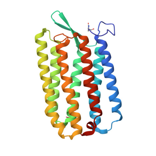
    6S63

  • PubMed Abstract: 

    Room-temperature diffraction methods are highly desirable for dynamic studies of biological macromolecules, since they allow high-resolution structural data to be collected as proteins undergo conformational changes. For crystals grown in lipidic cubic phase (LCP), an extruder is commonly used to pass a stream of microcrystals through the X-ray beam; however, the sample quantities required for this method may be difficult to produce for many membrane proteins. A more sample-efficient environment was created using two layers of low X-ray transmittance polymer films to mount crystals of the archaerhodopsin-3 (AR3) photoreceptor and room-temperature diffraction data were acquired. By using transparent and opaque polymer films, two structures, one corresponding to the desensitized, dark-adapted (DA) state and the other to the ground or light-adapted (LA) state, were solved to better than 1.9 Å resolution. All of the key structural features of AR3 were resolved, including the retinal chromophore, which is present as the 13-cis isomer in the DA state and as the all-trans isomer in the LA state. The film-sandwich sample environment enables diffraction data to be recorded at room temperature in both illuminated and dark conditions, which more closely approximate those in vivo. This simple approach is applicable to a wide range of membrane proteins crystallized in LCP and light-sensitive samples in general at synchrotron and laboratory X-ray sources.


  • Organizational Affiliation

    Diamond Light Source, Harwell Science and Innovation Campus, Didcot OX11 0DE, United Kingdom.


Macromolecules
Find similar proteins by:  (by identity cutoff)  |  3D Structure
Entity ID: 1
MoleculeChains Sequence LengthOrganismDetailsImage
Archaerhodopsin-3241Halorubrum sodomenseMutation(s): 1 
Membrane Entity: Yes 
UniProt
Find proteins for P96787 (Halorubrum sodomense)
Explore P96787 
Go to UniProtKB:  P96787
Entity Groups  
Sequence Clusters30% Identity50% Identity70% Identity90% Identity95% Identity100% Identity
UniProt GroupP96787
Sequence Annotations
Expand
  • Reference Sequence
Small Molecules
Ligands 6 Unique
IDChains Name / Formula / InChI Key2D Diagram3D Interactions
RET (Subject of Investigation/LOI)
Query on RET

Download Ideal Coordinates CCD File 
B [auth A]RETINAL
C20 H28 O
NCYCYZXNIZJOKI-OVSJKPMPSA-N
R16
Query on R16

Download Ideal Coordinates CCD File 
C [auth A],
D [auth A],
E [auth A],
F [auth A]
HEXADECANE
C16 H34
DCAYPVUWAIABOU-UHFFFAOYSA-N
DD9
Query on DD9

Download Ideal Coordinates CCD File 
G [auth A],
H [auth A],
I [auth A],
J [auth A],
K [auth A]
nonane
C9 H20
BKIMMITUMNQMOS-UHFFFAOYSA-N
CA
Query on CA

Download Ideal Coordinates CCD File 
L [auth A]CALCIUM ION
Ca
BHPQYMZQTOCNFJ-UHFFFAOYSA-N
CL
Query on CL

Download Ideal Coordinates CCD File 
N [auth A],
O [auth A],
P [auth A]
CHLORIDE ION
Cl
VEXZGXHMUGYJMC-UHFFFAOYSA-M
NA
Query on NA

Download Ideal Coordinates CCD File 
M [auth A]SODIUM ION
Na
FKNQFGJONOIPTF-UHFFFAOYSA-N
Modified Residues  1 Unique
IDChains TypeFormula2D DiagramParent
PCA
Query on PCA
A
L-PEPTIDE LINKINGC5 H7 N O3GLN
Experimental Data & Validation

Experimental Data

  • Method: X-RAY DIFFRACTION
  • Resolution: 1.85 Å
  • R-Value Free: 0.192 
  • R-Value Work: 0.175 
  • R-Value Observed: 0.176 
  • Space Group: P 21 21 21
Unit Cell:
Length ( Å )Angle ( ˚ )
a = 45.91α = 90
b = 48.351β = 90
c = 105.364γ = 90
Software Package:
Software NamePurpose
REFMACrefinement
DIALSdata reduction
Aimlessdata scaling
PHASERphasing
PDB_EXTRACTdata extraction

Structure Validation

View Full Validation Report


Ligand Structure Quality Assessment 


Entry History & Funding Information

Deposition Data


Funding OrganizationLocationGrant Number
Other governmentUnited Kingdom--
Biotechnology and Biological Sciences Research CouncilUnited KingdomBB/N006011/1
Wellcome TrustUnited Kingdom202892/Z/16/Z

Revision History  (Full details and data files)

  • Version 1.0: 2020-07-22
    Type: Initial release
  • Version 1.1: 2022-01-19
    Changes: Database references, Derived calculations
  • Version 1.2: 2024-01-24
    Changes: Data collection, Refinement description