6S36

Crystal structure of E. coli Adenylate kinase R119K mutant


Experimental Data Snapshot

  • Method: X-RAY DIFFRACTION
  • Resolution: 1.60 Å
  • R-Value Free: 0.236 
  • R-Value Work: 0.159 
  • R-Value Observed: 0.163 

wwPDB Validation   3D Report Full Report


This is version 1.2 of the entry. See complete history


Literature

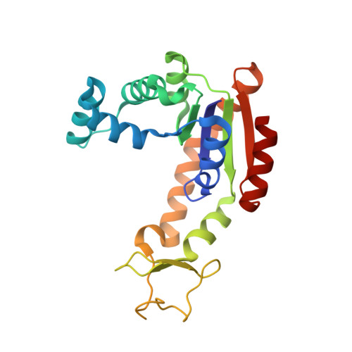
Nucleation of an Activating Conformational Change by a Cation-pi Interaction.

Rogne, P.Andersson, D.Grundstrom, C.Sauer-Eriksson, E.Linusson, A.Wolf-Watz, M.

(2019) Biochemistry 58: 3408-3412

  • DOI: https://doi.org/10.1021/acs.biochem.9b00538
  • Primary Citation of Related Structures:  
    6RZE, 6S36

  • PubMed Abstract: 

    As a key molecule in biology, adenosine triphosphate (ATP) has numerous crucial functions in, for instance, energetics, post-translational modifications, nucleotide biosynthesis, and cofactor metabolism. Here, we have discovered an intricate interplay between the enzyme adenylate kinase and its substrate ATP. The side chain of an arginine residue was found to be an efficient sensor of the aromatic moiety of ATP through the formation of a strong cation-π interaction. In addition to recognition, the interaction was found to have dual functionality. First, it nucleates the activating conformational transition of the ATP binding domain and also affects the specificity in the distant AMP binding domain. In light of the functional consequences resulting from the cation-π interaction, it is possible that the mode of ATP recognition may be a useful tool in enzyme design.


  • Organizational Affiliation

    Department of Chemistry , Umeå University , SE-901 87 Umeå , Sweden.


Macromolecules
Find similar proteins by:  (by identity cutoff)  |  3D Structure
Entity ID: 1
MoleculeChains Sequence LengthOrganismDetailsImage
Adenylate kinase214Escherichia coliMutation(s): 0 
Gene Names: 
EC: 2.7.4.3
UniProt
Find proteins for P69441 (Escherichia coli (strain K12))
Explore P69441 
Go to UniProtKB:  P69441
Entity Groups  
Sequence Clusters30% Identity50% Identity70% Identity90% Identity95% Identity100% Identity
UniProt GroupP69441
Sequence Annotations
Expand
  • Reference Sequence
Experimental Data & Validation

Experimental Data

  • Method: X-RAY DIFFRACTION
  • Resolution: 1.60 Å
  • R-Value Free: 0.236 
  • R-Value Work: 0.159 
  • R-Value Observed: 0.163 
  • Space Group: C 1 2 1
Unit Cell:
Length ( Å )Angle ( ˚ )
a = 135.69α = 90
b = 31.619β = 111.86
c = 53.171γ = 90
Software Package:
Software NamePurpose
PHENIXrefinement
XDSdata reduction
Aimlessdata scaling
PHASERphasing

Structure Validation

View Full Validation Report


Entry History & Funding Information

Deposition Data


Funding OrganizationLocationGrant Number
Swedish Research CouncilSweden--

Revision History  (Full details and data files)

  • Version 1.0: 2019-08-07
    Type: Initial release
  • Version 1.1: 2019-08-21
    Changes: Data collection, Database references
  • Version 1.2: 2024-01-24
    Changes: Data collection, Database references, Derived calculations, Refinement description