6S32

Crystal structure of ene-reductase CtOYE from Chroococcidiopsis thermalis.


Experimental Data Snapshot

  • Method: X-RAY DIFFRACTION
  • Resolution: 1.35 Å
  • R-Value Free: 0.255 
  • R-Value Work: 0.206 
  • R-Value Observed: 0.208 

wwPDB Validation   3D Report Full Report


Ligand Structure Quality Assessment 


This is version 2.1 of the entry. See complete history


Literature

Two new ene-reductases from photosynthetic extremophiles enlarge the panel of old yellow enzymes: CtOYE and GsOYE.

Robescu, M.S.Niero, M.Hall, M.Cendron, L.Bergantino, E.

(2020) Appl Microbiol Biotechnol 104: 2051-2066

  • DOI: https://doi.org/10.1007/s00253-019-10287-2
  • Primary Citation of Related Structures:  
    6S0G, 6S23, 6S31, 6S32

  • PubMed Abstract: 

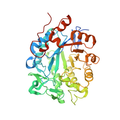
    Looking for new ene-reductases with uncovered features beneficial for biotechnological applications, by mining genomes of photosynthetic extremophile organisms, we identified two new Old Yellow Enzyme homologues: CtOYE, deriving from the cyanobacterium Chroococcidiopsis thermalis, and GsOYE, from the alga Galdieria sulphuraria. Both enzymes were produced and purified with very good yields and displayed catalytic activity on a broad substrate spectrum by reducing α,β-unsaturated ketones, aldehydes, maleimides and nitroalkenes with good to excellent stereoselectivity. Both enzymes prefer NADPH but demonstrate a good acceptance of NADH as cofactor. CtOYE and GsOYE represent robust biocatalysts showing high thermostability, a wide range of pH optimum and good co-solvent tolerance. High resolution X-ray crystal structures of both enzymes have been determined, revealing conserved features of the classical OYE subfamily as well as unique properties, such as a very long loop entering the active site or an additional C-terminal alpha helix in GsOYE. Not surprisingly, the active site of CtOYE and GsOYE structures revealed high affinity toward anions caught from the mother liquor and trapped in the anion hole where electron-withdrawing groups such as carbonyl group are engaged. Ligands (para-hydroxybenzaldehyde and 2-methyl-cyclopenten-1-one) added on purpose to study complexes of GsOYE were detected in the enzyme catalytic cavity, stacking on top of the FMN cofactor, and support the key role of conserved residues and FMN cofactor in the catalysis.


  • Organizational Affiliation

    Department of Biology, University of Padova, Viale G. Colombo 3, 35131, Padova, Italy.


Macromolecules
Find similar proteins by:  (by identity cutoff)  |  3D Structure
Entity ID: 1
MoleculeChains Sequence LengthOrganismDetailsImage
NADH:flavin oxidoreductase/NADH oxidase
A, B, C, D
390Chroococcidiopsis thermalisMutation(s): 0 
Gene Names: Chro_0590
UniProt
Find proteins for K9TVC9 (Chroococcidiopsis thermalis (strain PCC 7203))
Explore K9TVC9 
Go to UniProtKB:  K9TVC9
Entity Groups  
Sequence Clusters30% Identity50% Identity70% Identity90% Identity95% Identity100% Identity
UniProt GroupK9TVC9
Sequence Annotations
Expand
  • Reference Sequence
Small Molecules
Ligands 3 Unique
IDChains Name / Formula / InChI Key2D Diagram3D Interactions
FMN (Subject of Investigation/LOI)
Query on FMN

Download Ideal Coordinates CCD File 
G [auth A],
I [auth B],
K [auth C],
M [auth D]
FLAVIN MONONUCLEOTIDE
C17 H21 N4 O9 P
FVTCRASFADXXNN-SCRDCRAPSA-N
BEN
Query on BEN

Download Ideal Coordinates CCD File 
F [auth A]BENZAMIDINE
C7 H8 N2
PXXJHWLDUBFPOL-UHFFFAOYSA-N
ACT
Query on ACT

Download Ideal Coordinates CCD File 
E [auth A],
H [auth B],
J [auth C],
L [auth D]
ACETATE ION
C2 H3 O2
QTBSBXVTEAMEQO-UHFFFAOYSA-M
Experimental Data & Validation

Experimental Data

  • Method: X-RAY DIFFRACTION
  • Resolution: 1.35 Å
  • R-Value Free: 0.255 
  • R-Value Work: 0.206 
  • R-Value Observed: 0.208 
  • Space Group: P 1
Unit Cell:
Length ( Å )Angle ( ˚ )
a = 49.658α = 104.23
b = 78.172β = 99.98
c = 108.685γ = 90.04
Software Package:
Software NamePurpose
REFMACrefinement
XDSdata reduction
Aimlessdata scaling
PHASERphasing

Structure Validation

View Full Validation Report


Ligand Structure Quality Assessment 


Entry History 

Deposition Data

Revision History  (Full details and data files)

  • Version 1.0: 2020-01-29
    Type: Initial release
  • Version 1.1: 2020-02-19
    Changes: Database references
  • Version 2.0: 2021-08-04
    Changes: Atomic model, Data collection, Derived calculations, Non-polymer description, Refinement description, Structure summary
  • Version 2.1: 2024-01-24
    Changes: Data collection, Database references, Refinement description