6RXF

Crystal Structure of Bifidobacterium longum Multiple Inositol Polyphosphate Phosphatase Phosphohistidine Intermediate


Experimental Data Snapshot

  • Method: X-RAY DIFFRACTION
  • Resolution: 2.40 Å
  • R-Value Free: 0.235 
  • R-Value Work: 0.182 

wwPDB Validation   3D Report Full Report


This is version 1.3 of the entry. See complete history


Literature

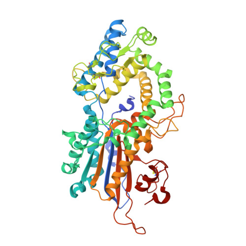
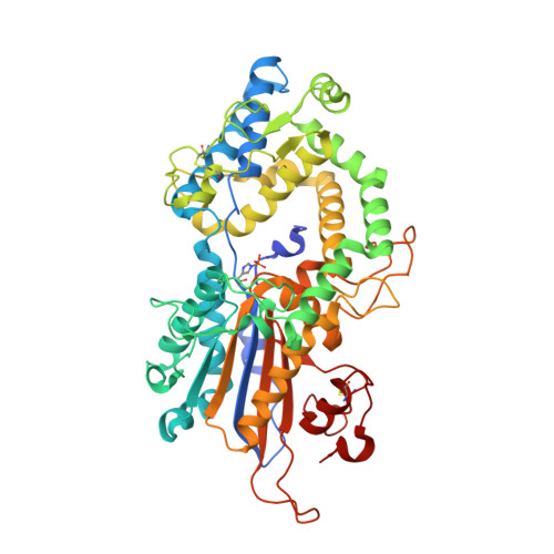
Snapshots during the catalytic cycle of a histidine acid phytase reveal an induced-fit structural mechanism.

Acquistapace, I.M.Zi Etek, M.A.Li, A.W.H.Salmon, M.Kuhn, I.Bedford, M.R.Brearley, C.A.Hemmings, A.M.

(2020) J Biol Chem 295: 17724-17737

  • DOI: https://doi.org/10.1074/jbc.RA120.015925
  • Primary Citation of Related Structures:  
    6RXD, 6RXE, 6RXF, 6RXG

  • PubMed Abstract: 

    Highly engineered phytases, which sequentially hydrolyze the hexakisphosphate ester of inositol known as phytic acid, are routinely added to the feeds of monogastric animals to improve phosphate bioavailability. New phytases are sought as starting points to further optimize the rate and extent of dephosphorylation of phytate in the animal digestive tract. Multiple inositol polyphosphate phosphatases (MINPPs) are clade 2 histidine phosphatases (HP2P) able to carry out the stepwise hydrolysis of phytate. MINPPs are not restricted by a strong positional specificity making them attractive targets for development as feed enzymes. Here, we describe the characterization of a MINPP from the Gram-positive bacterium Bifidobacterium longum (BlMINPP). BlMINPP has a typical HP2P-fold but, unusually, possesses a large α-domain polypeptide insertion relative to other MINPPs. This insertion, termed the U-loop, spans the active site and contributes to substrate specificity pockets underpopulated in other HP2Ps. Mutagenesis of U-loop residues reveals its contribution to enzyme kinetics and thermostability. Moreover, four crystal structures of the protein along the catalytic cycle capture, for the first time in an HP2P, a large ligand-driven α-domain motion essential to allow substrate access to the active site. This motion recruits residues both downstream of a molecular hinge and on the U-loop to participate in specificity subsites, and mutagenesis identified a mobile lysine residue as a key determinant of positional specificity of the enzyme. Taken together, these data provide important new insights to the factors determining stability, substrate recognition, and the structural mechanism of hydrolysis in this industrially important group of enzymes.


  • Organizational Affiliation

    School of Biological Sciences, University of East Anglia, Norwich, United Kingdom.


Macromolecules
Find similar proteins by:  (by identity cutoff)  |  3D Structure
Entity ID: 1
MoleculeChains Sequence LengthOrganismDetailsImage
Histidine acid phosphatase519Bifidobacterium longum subsp. infantis ATCC 15697 = JCM 1222 = DSM 20088Mutation(s): 1 
Gene Names: Blon_0263
UniProt
Find proteins for B7GTV0 (Bifidobacterium longum subsp. infantis (strain ATCC 15697 / DSM 20088 / JCM 1222 / NCTC 11817 / S12))
Explore B7GTV0 
Go to UniProtKB:  B7GTV0
Entity Groups  
Sequence Clusters30% Identity50% Identity70% Identity90% Identity95% Identity100% Identity
UniProt GroupB7GTV0
Sequence Annotations
Expand
  • Reference Sequence
Find similar proteins by:  (by identity cutoff)  |  3D Structure
Entity ID: 2
MoleculeChains Sequence LengthOrganismDetailsImage
Histidine acid phosphatase519Bifidobacterium longum subsp. infantis ATCC 15697 = JCM 1222 = DSM 20088Mutation(s): 1 
Gene Names: Blon_0263
UniProt
Find proteins for B7GTV0 (Bifidobacterium longum subsp. infantis (strain ATCC 15697 / DSM 20088 / JCM 1222 / NCTC 11817 / S12))
Explore B7GTV0 
Go to UniProtKB:  B7GTV0
Entity Groups  
Sequence Clusters30% Identity50% Identity70% Identity90% Identity95% Identity100% Identity
UniProt GroupB7GTV0
Sequence Annotations
Expand
  • Reference Sequence
Small Molecules
Ligands 1 Unique
IDChains Name / Formula / InChI Key2D Diagram3D Interactions
ZN (Subject of Investigation/LOI)
Query on ZN

Download Ideal Coordinates CCD File 
C [auth A]
D [auth A]
E [auth A]
F [auth A]
G [auth A]
C [auth A],
D [auth A],
E [auth A],
F [auth A],
G [auth A],
H [auth A],
I [auth A],
J [auth A],
K [auth B],
L [auth B],
M [auth B],
N [auth B],
O [auth B],
P [auth B],
Q [auth B],
R [auth B],
S [auth B]
ZINC ION
Zn
PTFCDOFLOPIGGS-UHFFFAOYSA-N
Modified Residues  1 Unique
IDChains TypeFormula2D DiagramParent
NEP
Query on NEP
B
L-PEPTIDE LINKINGC6 H10 N3 O5 PHIS
Experimental Data & Validation

Experimental Data

  • Method: X-RAY DIFFRACTION
  • Resolution: 2.40 Å
  • R-Value Free: 0.235 
  • R-Value Work: 0.182 
  • Space Group: P 1
Unit Cell:
Length ( Å )Angle ( ˚ )
a = 55.508α = 71.3
b = 73.118β = 72.99
c = 88.987γ = 78.99
Software Package:
Software NamePurpose
PHENIXrefinement
xia2data reduction
xia2data scaling
PHASERphasing

Structure Validation

View Full Validation Report


Entry History & Funding Information

Deposition Data


Funding OrganizationLocationGrant Number
Biotechnology and Biological Sciences Research CouncilUnited KingdomBB/M022978/1

Revision History  (Full details and data files)

  • Version 1.0: 2020-07-08
    Type: Initial release
  • Version 1.1: 2021-01-27
    Changes: Database references, Derived calculations
  • Version 1.2: 2021-02-24
    Changes: Database references
  • Version 1.3: 2024-01-24
    Changes: Advisory, Data collection, Database references, Refinement description