6RU4

Structure of the SBP FpvC from pseudomonas aeruginosa in complex with Mn2+


Experimental Data Snapshot

  • Method: X-RAY DIFFRACTION
  • Resolution: 2.49 Å
  • R-Value Free: 0.238 
  • R-Value Work: 0.196 
  • R-Value Observed: 0.198 

wwPDB Validation   3D Report Full Report


Ligand Structure Quality Assessment 


This is version 1.3 of the entry. See complete history


Literature

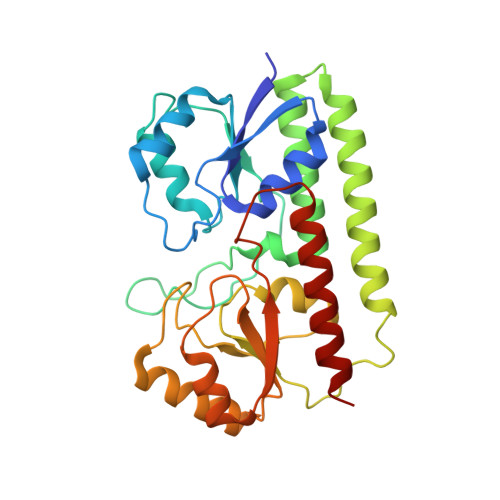
A unique ferrous iron binding mode is associated with large conformational changes for the transport protein FpvC of Pseudomonas aeruginosa.

Vigouroux, A.Aumont-Nicaise, M.Boussac, A.Marty, L.Lo Bello, L.Legrand, P.Brillet, K.Schalk, I.J.Morera, S.

(2020) FEBS J 287: 295-309

  • DOI: https://doi.org/10.1111/febs.15004
  • Primary Citation of Related Structures:  
    6R3Z, 6R44, 6R5S, 6R6K, 6RU4

  • PubMed Abstract: 

    Pseudomonas aeruginosa secretes pyoverdine, a major siderophore to get access to iron, an essential nutrient. Pyoverdine scavenges ferric iron in the bacterial environment with the resulting complex internalized by bacteria. Releasing of iron from pyoverdine in the periplasm involves an iron reduction by an inner membrane reductase and two solute-binding proteins (SBPs) FpvC and FpvF in association with their ABC transporter. FpvC and FpvF belong to two different subgroups of SBPs within the structural cluster A: FpvC and FpvF were proposed to be a metal-binding protein and a ferrisiderophore-binding protein respectively. Here, we report the redox state and the binding mode of iron to FpvC. We first solved the crystal structure of FpvC bound to a fortuitous Ni 2+ by single anomalous dispersion method. Using a different protein purification strategy, we determined the structure of FpvC with manganese and iron, which binds to FpvC in a ferrous state as demonstrated by electron paramagnetic resonance. FpvC is the first example of a hexahistidine metal site among SBPs in which the Fe 2+ redox state is stabilized under aerobic conditions. Using biophysics methods, we showed that FpvC reversibly bind to a broad range of divalent ions. The structure of a mutant mimicking the apo FpvC reveals a protein in an open state with large conformational changes when compared with the metal-bound FpvC. These results highlight that the canonical metal site in FpvC is distinct from those yet described in SBPs and they provide new insights into the mechanism of PVD-Fe dissociation in P. aeruginosa.


  • Organizational Affiliation

    Institute for Integrative Biology of the Cell (I2BC), CNRS CEA Univ. Paris-Sud, Université Paris-Saclay, Gif-sur-Yvette, France.


Macromolecules
Find similar proteins by:  (by identity cutoff)  |  3D Structure
Entity ID: 1
MoleculeChains Sequence LengthOrganismDetailsImage
Probable adhesion protein
A, B, C, D
281Pseudomonas aeruginosa PAO1Mutation(s): 0 
Gene Names: PA2407
UniProt
Find proteins for Q9I174 (Pseudomonas aeruginosa (strain ATCC 15692 / DSM 22644 / CIP 104116 / JCM 14847 / LMG 12228 / 1C / PRS 101 / PAO1))
Explore Q9I174 
Go to UniProtKB:  Q9I174
Entity Groups  
Sequence Clusters30% Identity50% Identity70% Identity90% Identity95% Identity100% Identity
UniProt GroupQ9I174
Sequence Annotations
Expand
  • Reference Sequence
Small Molecules
Ligands 3 Unique
IDChains Name / Formula / InChI Key2D Diagram3D Interactions
PEG (Subject of Investigation/LOI)
Query on PEG

Download Ideal Coordinates CCD File 
R [auth A],
S [auth A]
DI(HYDROXYETHYL)ETHER
C4 H10 O3
MTHSVFCYNBDYFN-UHFFFAOYSA-N
EDO (Subject of Investigation/LOI)
Query on EDO

Download Ideal Coordinates CCD File 
AA [auth B]
NA [auth D]
OA [auth D]
Q [auth A]
X [auth B]
AA [auth B],
NA [auth D],
OA [auth D],
Q [auth A],
X [auth B],
Y [auth B],
Z [auth B]
1,2-ETHANEDIOL
C2 H6 O2
LYCAIKOWRPUZTN-UHFFFAOYSA-N
MN (Subject of Investigation/LOI)
Query on MN

Download Ideal Coordinates CCD File 
BA [auth C]
CA [auth C]
DA [auth C]
E [auth A]
EA [auth C]
BA [auth C],
CA [auth C],
DA [auth C],
E [auth A],
EA [auth C],
F [auth A],
FA [auth C],
G [auth A],
GA [auth C],
H [auth A],
HA [auth C],
I [auth A],
IA [auth C],
J [auth A],
JA [auth C],
K [auth A],
KA [auth D],
L [auth A],
LA [auth D],
M [auth A],
MA [auth D],
N [auth A],
O [auth A],
P [auth A],
T [auth B],
U [auth B],
V [auth B],
W [auth B]
MANGANESE (II) ION
Mn
WAEMQWOKJMHJLA-UHFFFAOYSA-N
Experimental Data & Validation

Experimental Data

  • Method: X-RAY DIFFRACTION
  • Resolution: 2.49 Å
  • R-Value Free: 0.238 
  • R-Value Work: 0.196 
  • R-Value Observed: 0.198 
  • Space Group: P 1 21 1
Unit Cell:
Length ( Å )Angle ( ˚ )
a = 44.01α = 90
b = 178.29β = 90.61
c = 66.82γ = 90
Software Package:
Software NamePurpose
BUSTERrefinement
XDSdata reduction
XSCALEdata scaling
PHASERphasing

Structure Validation

View Full Validation Report


Ligand Structure Quality Assessment 


Entry History 

Deposition Data

Revision History  (Full details and data files)

  • Version 1.0: 2019-07-31
    Type: Initial release
  • Version 1.1: 2019-08-07
    Changes: Data collection, Database references
  • Version 1.2: 2020-01-29
    Changes: Database references
  • Version 1.3: 2024-01-24
    Changes: Data collection, Database references, Refinement description